 Movie
Movie Controller

[English] 日本語
 Yorodumi
- PDB-1zt4: The crystal structure of human CD1d with and without alpha-Galact... -
+ Open data

- Basic information
| Entry | Database: PDB / ID: 1zt4 | |||||||||
|---|---|---|---|---|---|---|---|---|---|---|
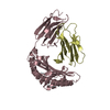
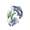
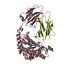
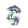
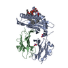
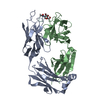
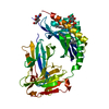
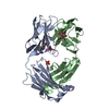
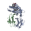
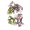
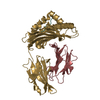
| Title | The crystal structure of human CD1d with and without alpha-Galactosylceramide | |||||||||
 Components Components |
| |||||||||
 Keywords Keywords |  IMMUNE SYSTEM / human CD1d / CD1 / MHC class I / empty binding groove / glycolipid / alpha-galactosylceramide / alpha-GalCer / Structural Proteomics in Europe / SPINE / Structural Genomics IMMUNE SYSTEM / human CD1d / CD1 / MHC class I / empty binding groove / glycolipid / alpha-galactosylceramide / alpha-GalCer / Structural Proteomics in Europe / SPINE / Structural Genomics | |||||||||
| Function / homology |  Function and homology information Function and homology informationlipid antigen binding / T cell selection / endogenous lipid antigen binding / exogenous lipid antigen binding / antigen processing and presentation, endogenous lipid antigen via MHC class Ib / antigen processing and presentation, exogenous lipid antigen via MHC class Ib / lipopeptide binding / positive regulation of innate immune response / heterotypic cell-cell adhesion / beta-2-microglobulin binding ...lipid antigen binding / T cell selection / endogenous lipid antigen binding / exogenous lipid antigen binding / antigen processing and presentation, endogenous lipid antigen via MHC class Ib / antigen processing and presentation, exogenous lipid antigen via MHC class Ib / lipopeptide binding / positive regulation of innate immune response / heterotypic cell-cell adhesion / beta-2-microglobulin binding / positive regulation of T cell proliferation / detection of bacterium / cell adhesion molecule binding / positive regulation of ferrous iron binding / positive regulation of transferrin receptor binding / early endosome lumen / positive regulation of receptor binding / Nef mediated downregulation of MHC class I complex cell surface expression / DAP12 interactions / negative regulation of receptor binding / Endosomal/Vacuolar pathway / Antigen Presentation: Folding, assembly and peptide loading of class I MHC / antigen processing and presentation of exogenous protein antigen via MHC class Ib, TAP-dependent / cellular response to iron(III) ion / negative regulation of forebrain neuron differentiation / ER to Golgi transport vesicle membrane / response to molecule of bacterial origin / regulation of erythrocyte differentiation / regulation of iron ion transport / MHC class I peptide loading complex / HFE-transferrin receptor complex / T cell mediated cytotoxicity / cellular response to iron ion / antigen processing and presentation of endogenous peptide antigen via MHC class I / positive regulation of T cell cytokine production / MHC class I protein complex / multicellular organismal-level iron ion homeostasis / positive regulation of T cell mediated cytotoxicity / peptide antigen assembly with MHC class II protein complex / negative regulation of neurogenesis / MHC class II protein complex / positive regulation of receptor-mediated endocytosis / cellular response to nicotine / recycling endosome membrane / phagocytic vesicle membrane / specific granule lumen / peptide antigen binding / positive regulation of cellular senescence / antigen processing and presentation of exogenous peptide antigen via MHC class II / Immunoregulatory interactions between a Lymphoid and a non-Lymphoid cell / Interferon gamma signaling / positive regulation of immune response / negative regulation of epithelial cell proliferation / Modulation by Mtb of host immune system / positive regulation of T cell activation / sensory perception of smell / negative regulation of neuron projection development / tertiary granule lumen / DAP12 signaling / MHC class II protein complex binding / late endosome membrane / T cell differentiation in thymus / positive regulation of protein binding / ER-Phagosome pathway / iron ion transport / histone binding / protein refolding / early endosome membrane / protein homotetramerization / basolateral plasma membrane / intracellular iron ion homeostasis / amyloid fibril formation / lysosome / learning or memory / endosome membrane / immune response / Amyloid fiber formation / lysosomal membrane / endoplasmic reticulum lumen / external side of plasma membrane / Golgi membrane / focal adhesion / innate immune response / Neutrophil degranulation / endoplasmic reticulum membrane / SARS-CoV-2 activates/modulates innate and adaptive immune responses / Golgi apparatus / cell surface / endoplasmic reticulum / protein homodimerization activity / extracellular space / extracellular exosome / extracellular region / membrane / identical protein binding / plasma membrane / cytosol / cytoplasmSimilarity search - Function | |||||||||
| Biological species |  Homo sapiens (human) Homo sapiens (human) | |||||||||
| Method | X-RAY DIFFRACTION / SYNCHROTRON / MOLECULAR REPLACEMENT / Resolution: 3 Å | |||||||||
 Authors Authors | Koch, M. / Stronge, V.S. / Shepherd, D. / Gadola, S.D. / Mathew, B. / Ritter, G. / Fersht, A.R. / Besra, G.S. / Schmidt, R.R. / Jones, E.Y. ...Koch, M. / Stronge, V.S. / Shepherd, D. / Gadola, S.D. / Mathew, B. / Ritter, G. / Fersht, A.R. / Besra, G.S. / Schmidt, R.R. / Jones, E.Y. / Cerundolo, V. / Structural Proteomics in Europe (SPINE) | |||||||||
 Citation Citation | Journal: Nat.Immunol. / Year: 2005 Title: The crystal structure of human CD1d with and without alpha-galactosylceramide Authors: Koch, M. / Stronge, V.S. / Shepherd, D. / Gadola, S.D. / Mathew, B. / Ritter, G. / Fersht, A.R. / Besra, G.S. / Schmidt, R.R. / Jones, E.Y. / Cerundolo, V. | |||||||||
| History |
|
- Structure visualization
| Structure viewer | Molecule: MolmilJmol/JSmol |
|---|
- Downloads & links
-Download
| PDBx/mmCIF format | 1zt4.cif.gz | 162.9 KB | Display | PDBx/mmCIF format |
|---|---|---|---|---|
| PDB format | pdb1zt4.ent.gz | 128.6 KB | Display | PDB format |
| PDBx/mmJSON format | 1zt4.json.gz | Tree view | PDBx/mmJSON format | |
| Others |  Other downloads Other downloads |
-Validation report
| Arichive directory | https://data.pdbj.org/pub/pdb/validation_reports/zt/1zt4ftp://data.pdbj.org/pub/pdb/validation_reports/zt/1zt4 | HTTPS FTP |
|---|
-Related structure data
| Related structure data |  1cd1S S: Starting model for refinement |
|---|---|
| Similar structure data | |
| Other databases |


-Links
 PDBj
PDBj

- Assembly
| Deposited unit | 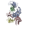
| ||||||||
|---|---|---|---|---|---|---|---|---|---|
| 1 | 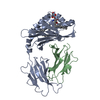
| ||||||||
| 2 | 
| ||||||||
| 3 |
| ||||||||
| Unit cell |
| ||||||||
| Details | the biological assembly is heavy chain from CD1d plus beta-microglobulin, chains A and B (ligand bound molecule) or chains C and D (non-ligand or empty molecule). |
-Components
| #1: Protein | Mass: 31818.814 Da / Num. of mol.: 2 / Fragment: CD1d heavy chain Source method: isolated from a genetically manipulated source Source: (gene. exp.) Homo sapiens (human) / Gene: CD1D / Plasmid: pET23d / Production host:  Escherichia coli (E. coli) / Strain (production host): HMS174 / References: UniProt: P15813 Escherichia coli (E. coli) / Strain (production host): HMS174 / References: UniProt: P15813#2: Protein | Beta-2 microglobulin / HDCMA22PMass: 11879.356 Da / Num. of mol.: 2 Source method: isolated from a genetically manipulated source Source: (gene. exp.) Homo sapiens (human) / Gene: B2M / Plasmid: pET23d / Production host: Escherichia coli (E. coli) / Strain (production host): HMS174 / References: UniProt: P61769#3: Sugar | ChemComp-AGH / | Alpha-Galactosylceramide  Type: D-saccharide / Mass: 858.322 Da / Num. of mol.: 1 Type: D-saccharide / Mass: 858.322 Da / Num. of mol.: 1Source method: isolated from a genetically manipulated source Formula: C50H99NO9 #4: Water | ChemComp-HOH / | Water Mass: 18.015 Da / Num. of mol.: 18 / Source method: isolated from a natural source / Formula: H2O Mass: 18.015 Da / Num. of mol.: 18 / Source method: isolated from a natural source / Formula: H2O |
|---|
-Experimental details
-Experiment
| Experiment | Method: X-RAY DIFFRACTION / Number of used crystals: 1 |
|---|
- Sample preparation
| Crystal | Density Matthews: 2.6 Å3/Da / Density % sol: 53 % |
|---|---|
| Crystal grow | Temperature: 293 K / Method: vapor diffusion, sitting drop / pH: 7.5 Details: PEG3350, potassium fluoride, pH 7.5, VAPOR DIFFUSION, SITTING DROP, temperature 293.0K |
-Data collection
| Diffraction | Mean temperature: 100 K |
|---|---|
| Diffraction source | Source: SYNCHROTRON / Site: ESRF  / Beamline: ID14-1 / Wavelength: 0.934 Å / Beamline: ID14-1 / Wavelength: 0.934 Å |
| Detector | Type: ADSC QUANTUM 4 / Detector: CCD / Date: Oct 11, 2004 |
| Radiation | Protocol: SINGLE WAVELENGTH / Monochromatic (M) / Laue (L): M / Scattering type: x-ray |
| Radiation wavelength | Wavelength: 0.934 Å / Relative weight: 1 |
| Reflection | Resolution: 3→30 Å / Num. obs: 17523 / % possible obs: 99.8 % / Observed criterion σ(F): 0.5 / Observed criterion σ(I): 0.5 / Redundancy: 11.2 % / Rmerge(I) obs: 0.184 / Rsym value: 0.184 / Net I/σ(I): 15.2 |
| Reflection shell | Resolution: 3→3.19 Å / Redundancy: 11.2 % / Rmerge(I) obs: 0.94 / Mean I/σ(I) obs: 3.2 / Num. unique all: 2864 / Rsym value: 0.94 / % possible all: 100 |
- Processing
| Software |
| ||||||||||||||||||||||||||||
|---|---|---|---|---|---|---|---|---|---|---|---|---|---|---|---|---|---|---|---|---|---|---|---|---|---|---|---|---|---|
| Refinement | Method to determine structure: MOLECULAR REPLACEMENT Starting model: PDB ENTRY 1CD1 Resolution: 3→30 Å / Isotropic thermal model: isotropic / Cross valid method: THROUGHOUT / σ(F): 0 / Stereochemistry target values: Engh & Huber
| ||||||||||||||||||||||||||||
| Solvent computation | Bsol: 80 Å2 | ||||||||||||||||||||||||||||
| Displacement parameters | Biso mean: 32.247 Å2
| ||||||||||||||||||||||||||||
| Refinement step | Cycle: LAST / Resolution: 3→30 Å
| ||||||||||||||||||||||||||||
| Refine LS restraints |
| ||||||||||||||||||||||||||||
| LS refinement shell | Resolution: 3→3.14 Å
| ||||||||||||||||||||||||||||
| Xplor file |
|
