 Movie
Movie Controller


+ Open data

- Basic information
| Entry | Database: PDB / ID: 1z9m | ||||||
|---|---|---|---|---|---|---|---|
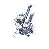
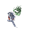
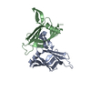
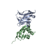
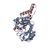
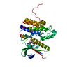
| Title | Crystal Structure of Nectin-like molecule-1 protein Domain 1 | ||||||
 Components Components | GAPA225 | ||||||
 Keywords Keywords |  CELL ADHESION / Nectin-like / Ig-like domain / V domain CELL ADHESION / Nectin-like / Ig-like domain / V domain | ||||||
| Function / homology |  Function and homology information Function and homology informationNectin/Necl trans heterodimerization / Adherens junctions interactions / adherens junction organization / heterophilic cell-cell adhesion via plasma membrane cell adhesion molecules / homophilic cell adhesion via plasma membrane adhesion molecules / cell-cell junction / membrane => GO:0016020 / protein homodimerization activity / plasma membraneSimilarity search - Function | ||||||
| Biological species |  Homo sapiens (human) Homo sapiens (human) | ||||||
| Method | X-RAY DIFFRACTION / SYNCHROTRON / MAD / Resolution: 2.4 Å | ||||||
 Authors Authors | Dong, X. / Xu, F. / Gong, Y. / Gao, J. / Lin, P. / Chen, T. / Peng, Y. / Qiang, B. / Yuan, J. / Peng, X. / Rao, Z. | ||||||
 Citation Citation | Journal: J.Biol.Chem. / Year: 2006 Title: Crystal Structure of the V Domain of Human Nectin-like Molecule-1/Syncam3/Tsll1/Igsf4b, a Neural Tissue-specific Immunoglobulin-like Cell-Cell Adhesion Molecule Authors: Dong, X. / Xu, F. / Gong, Y. / Gao, J. / Lin, P. / Chen, T. / Peng, Y. / Qiang, B. / Yuan, J. / Peng, X. / Rao, Z. | ||||||
| History |
|
- Structure visualization
| Structure viewer | Molecule: MolmilJmol/JSmol |
|---|
- Downloads & links
-Download
| PDBx/mmCIF format | 1z9m.cif.gz | 53.5 KB | Display | PDBx/mmCIF format |
|---|---|---|---|---|
| PDB format | pdb1z9m.ent.gz | 42.2 KB | Display | PDB format |
| PDBx/mmJSON format | 1z9m.json.gz | Tree view | PDBx/mmJSON format | |
| Others |  Other downloads Other downloads |
-Validation report
| Arichive directory | https://data.pdbj.org/pub/pdb/validation_reports/z9/1z9mftp://data.pdbj.org/pub/pdb/validation_reports/z9/1z9m | HTTPS FTP |
|---|
-Related structure data
| Similar structure data |
|---|


-Links
 PDBj
PDBj


- Assembly
| Deposited unit | 
| ||||||||
|---|---|---|---|---|---|---|---|---|---|
| 1 |
| ||||||||
| Unit cell |
|
-Components
| #1: Protein | Mass: 15868.651 Da / Num. of mol.: 2 / Fragment: residues 37-141 Source method: isolated from a genetically manipulated source Source: (gene. exp.) Homo sapiens (human) / Plasmid: pET32a / Species (production host): Escherichia coli / Production host:  Escherichia coli BL21 (bacteria) / Strain (production host): BL21 / References: GenBank: 37181789, UniProt: Q8N126*PLUS Escherichia coli BL21 (bacteria) / Strain (production host): BL21 / References: GenBank: 37181789, UniProt: Q8N126*PLUS#2: Water | ChemComp-HOH / | Water Mass: 18.015 Da / Num. of mol.: 196 / Source method: isolated from a natural source / Formula: H2O Mass: 18.015 Da / Num. of mol.: 196 / Source method: isolated from a natural source / Formula: H2O |
|---|
-Experimental details
-Experiment
| Experiment | Method: X-RAY DIFFRACTION / Number of used crystals: 1 |
|---|
- Sample preparation
| Crystal | Density Matthews: 2.4 Å3/Da / Density % sol: 48.5 % / Description: the file contains Friedel pairs. |
|---|---|
| Crystal grow | Temperature: 289 K / Method: vapor diffusion, hanging drop / pH: 8 Details: Sodium Formate, pH 8.0, VAPOR DIFFUSION, HANGING DROP, temperature 289K |
-Data collection
| Diffraction | Mean temperature: 100 K | ||||||||||||
|---|---|---|---|---|---|---|---|---|---|---|---|---|---|
| Diffraction source | Source: SYNCHROTRON / Site: BSRF  / Beamline: 3W1A / Wavelength: 0.9000, 0.9788, 0.9793 / Beamline: 3W1A / Wavelength: 0.9000, 0.9788, 0.9793 | ||||||||||||
| Detector | Type: MARRESEARCH / Detector: CCD / Date: Jan 10, 2005 | ||||||||||||
| Radiation | Monochromator: GRAPHITE / Protocol: MAD / Monochromatic (M) / Laue (L): M / Scattering type: x-ray | ||||||||||||
| Radiation wavelength |
| ||||||||||||
| Reflection | Resolution: 2.4→50 Å / Num. all: 24704 / Num. obs: 24704 / % possible obs: 100 % / Observed criterion σ(F): -3 / Observed criterion σ(I): 0 | ||||||||||||
| Reflection shell | Resolution: 2.4→2.49 Å / % possible all: 100 |
- Processing
| Software |
| ||||||||||||||||||||
|---|---|---|---|---|---|---|---|---|---|---|---|---|---|---|---|---|---|---|---|---|---|
| Refinement | Method to determine structure: MAD / Resolution: 2.4→48.2 Å / σ(F): 0 / Stereochemistry target values: Engh & Huber / Details: the file contains Friedel pairs.
| ||||||||||||||||||||
| Refinement step | Cycle: LAST / Resolution: 2.4→48.2 Å
| ||||||||||||||||||||
| Refine LS restraints |
|
