 Movie
Movie Controller


+ Open data

- Basic information
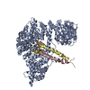
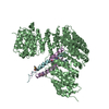
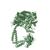
| Entry | Database: PDB / ID: 1z3h | ||||||
|---|---|---|---|---|---|---|---|
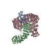
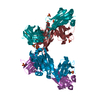
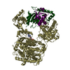
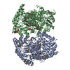
| Title | The exportin Cse1 in its cargo-free, cytoplasmic state | ||||||
 Components Components | Importin alpha re-exporter | ||||||
 Keywords Keywords |  PROTEIN TRANSPORT / Cse1 / Exportin / Nuclear transport / HEAT repeat PROTEIN TRANSPORT / Cse1 / Exportin / Nuclear transport / HEAT repeat | ||||||
| Function / homology |  Function and homology information Function and homology informationnuclear export signal receptor activity / snRNA import into nucleus / protein export from nucleus / positive regulation of protein export from nucleus / small GTPase binding / protein import into nucleus / nuclear envelope / cell cycle / cell division / protein-containing complex / cytosolSimilarity search - Function | ||||||
| Biological species |  Saccharomyces cerevisiae (brewer's yeast) Saccharomyces cerevisiae (brewer's yeast) | ||||||
| Method | X-RAY DIFFRACTION / SYNCHROTRON / SAD / Resolution: 3.1 Å | ||||||
 Authors Authors | Cook, A. / Fernandez, E. / Lindner, D. / Ebert, J. / Schlenstedt, G. / Conti, E. | ||||||
 Citation Citation | Journal: Mol.Cell / Year: 2005 Title: The structure of the nuclear export receptor cse1 in its cytosolic state reveals a closed conformation incompatible with cargo binding Authors: Cook, A. / Fernandez, E. / Lindner, D. / Ebert, J. / Schlenstedt, G. / Conti, E. | ||||||
| History |
|
- Structure visualization
| Structure viewer | Molecule: MolmilJmol/JSmol |
|---|
- Downloads & links
-Download
| PDBx/mmCIF format | 1z3h.cif.gz | 367 KB | Display | PDBx/mmCIF format |
|---|---|---|---|---|
| PDB format | pdb1z3h.ent.gz | 297.8 KB | Display | PDB format |
| PDBx/mmJSON format | 1z3h.json.gz | Tree view | PDBx/mmJSON format | |
| Others |  Other downloads Other downloads |
-Validation report
| Arichive directory | https://data.pdbj.org/pub/pdb/validation_reports/z3/1z3hftp://data.pdbj.org/pub/pdb/validation_reports/z3/1z3h | HTTPS FTP |
|---|
-Related structure data
| Related structure data | |
|---|---|
| Similar structure data |


-Links
 PDBj
PDBj

- Assembly
| Deposited unit | 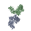
| ||||||||
|---|---|---|---|---|---|---|---|---|---|
| 1 | 
| ||||||||
| 2 | 
| ||||||||
| Unit cell |
|
-Components
| #1: Protein | Mass: 110526.672 Da / Num. of mol.: 2 Source method: isolated from a genetically manipulated source Source: (gene. exp.) Saccharomyces cerevisiae (brewer's yeast)Gene: Cse1 / Plasmid: PQE-60 / Production host:  Escherichia coli (E. coli) / Strain (production host): DL-41 / References: UniProt: P33307 Escherichia coli (E. coli) / Strain (production host): DL-41 / References: UniProt: P33307#2: Chemical | ChemComp-MG / |   Mass: 24.305 Da / Num. of mol.: 1 / Source method: obtained synthetically / Formula: Mg Mass: 24.305 Da / Num. of mol.: 1 / Source method: obtained synthetically / Formula: Mg |
|---|
-Experimental details
-Experiment
| Experiment | Method: X-RAY DIFFRACTION / Number of used crystals: 2 |
|---|
- Sample preparation
| Crystal | Density Matthews: 2.5 Å3/Da / Density % sol: 50.9 % |
|---|---|
| Crystal grow | Temperature: 298 K / Method: vapor diffusion, hanging drop / pH: 8 Details: PEG 8000, MgCl2, glycerol, DTT, pH 8.0, VAPOR DIFFUSION, HANGING DROP, temperature 298K |
-Data collection
| Diffraction |
| |||||||||||||||||||||||||||||||||||||||||||||||||||||||||||||||||||||||||||||
|---|---|---|---|---|---|---|---|---|---|---|---|---|---|---|---|---|---|---|---|---|---|---|---|---|---|---|---|---|---|---|---|---|---|---|---|---|---|---|---|---|---|---|---|---|---|---|---|---|---|---|---|---|---|---|---|---|---|---|---|---|---|---|---|---|---|---|---|---|---|---|---|---|---|---|---|---|---|---|
| Diffraction source |
| |||||||||||||||||||||||||||||||||||||||||||||||||||||||||||||||||||||||||||||
| Detector |
| |||||||||||||||||||||||||||||||||||||||||||||||||||||||||||||||||||||||||||||
| Radiation |
| |||||||||||||||||||||||||||||||||||||||||||||||||||||||||||||||||||||||||||||
| Radiation wavelength |
| |||||||||||||||||||||||||||||||||||||||||||||||||||||||||||||||||||||||||||||
| Reflection | Redundancy: 13.2 % / Av σ(I) over netI: 3.7 / Number: 33873 / Rmerge(I) obs: 0.102 / Rsym value: 0.102 / D res high: 3.3 Å / D res low: 122.06 Å / % possible obs: 98.6 | |||||||||||||||||||||||||||||||||||||||||||||||||||||||||||||||||||||||||||||
| Diffraction reflection shell |
| |||||||||||||||||||||||||||||||||||||||||||||||||||||||||||||||||||||||||||||
| Reflection | Resolution: 3.3→50 Å / Num. all: 44757 / Num. obs: 41661 / % possible obs: 98.5 % / Observed criterion σ(F): 1 / Observed criterion σ(I): 1 / Redundancy: 13.2 % / Biso Wilson estimate: 96.017 Å2 / Rsym value: 0.102 | |||||||||||||||||||||||||||||||||||||||||||||||||||||||||||||||||||||||||||||
| Reflection shell | Resolution: 3.3→3.48 Å / Redundancy: 13.6 % / Mean I/σ(I) obs: 5.5 / Rsym value: 0.391 / % possible all: 98.5 |


-Phasing
| Phasing | Method: SAD | |||||||||||||||||||||||||||||||||||||||||||||||||||||||||||||||||||||||||||||||||||||||||||||||||||||||||||||||||||||||||||||||||||||||||||||||||||||||||||||||||||||||||||||||||||||||||||||||||||||||||||||||||||||||||||||||||||||||||||||||||||||
|---|---|---|---|---|---|---|---|---|---|---|---|---|---|---|---|---|---|---|---|---|---|---|---|---|---|---|---|---|---|---|---|---|---|---|---|---|---|---|---|---|---|---|---|---|---|---|---|---|---|---|---|---|---|---|---|---|---|---|---|---|---|---|---|---|---|---|---|---|---|---|---|---|---|---|---|---|---|---|---|---|---|---|---|---|---|---|---|---|---|---|---|---|---|---|---|---|---|---|---|---|---|---|---|---|---|---|---|---|---|---|---|---|---|---|---|---|---|---|---|---|---|---|---|---|---|---|---|---|---|---|---|---|---|---|---|---|---|---|---|---|---|---|---|---|---|---|---|---|---|---|---|---|---|---|---|---|---|---|---|---|---|---|---|---|---|---|---|---|---|---|---|---|---|---|---|---|---|---|---|---|---|---|---|---|---|---|---|---|---|---|---|---|---|---|---|---|---|---|---|---|---|---|---|---|---|---|---|---|---|---|---|---|---|---|---|---|---|---|---|---|---|---|---|---|---|---|---|---|---|---|---|---|---|---|---|---|---|---|---|---|---|---|---|---|---|---|
| Phasing MAD set site |
|
- Processing
| Software |
| ||||||||||||||||||||||||||||
|---|---|---|---|---|---|---|---|---|---|---|---|---|---|---|---|---|---|---|---|---|---|---|---|---|---|---|---|---|---|
| Refinement | Method to determine structure: SAD / Resolution: 3.1→122 Å / σ(F): 0 / Stereochemistry target values: Engh & Huber Details: Residue side chains that were not visible in the final 2Fo-Fc map have been placed in the model but the B factors for these atoms have been set above 200. The protein was treated with ...Details: Residue side chains that were not visible in the final 2Fo-Fc map have been placed in the model but the B factors for these atoms have been set above 200. The protein was treated with subtilisin during purification. This resulted in two fragments of 100 kDa and 10 kDa respectively that co-purify and are both present in the crystal
| ||||||||||||||||||||||||||||
| Solvent computation | Bsol: 30.849 Å2 | ||||||||||||||||||||||||||||
| Displacement parameters | Biso mean: 77.326 Å2
| ||||||||||||||||||||||||||||
| Refinement step | Cycle: LAST / Resolution: 3.1→122 Å
| ||||||||||||||||||||||||||||
| Refine LS restraints |
| ||||||||||||||||||||||||||||
| LS refinement shell | Resolution: 3.1→3.2 Å
| ||||||||||||||||||||||||||||
| Xplor file |
|
