 Movie
Movie Controller


+ Open data

- Basic information
| Entry | Database: PDB / ID: 1ytw | ||||||
|---|---|---|---|---|---|---|---|
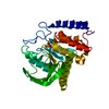
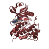
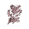
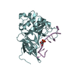
| Title | YERSINIA PTPASE COMPLEXED WITH TUNGSTATE | ||||||
 Components Components | YERSINIA PROTEIN TYROSINE PHOSPHATASE | ||||||
 Keywords Keywords | HYDROLASE / PROTEIN TYROSINE PHOSPHATASE | ||||||
| Function / homology |  Function and homology informationdephosphorylation / protein-tyrosine-phosphatase / protein tyrosine phosphatase activity / extracellular region Function and homology informationdephosphorylation / protein-tyrosine-phosphatase / protein tyrosine phosphatase activity / extracellular regionSimilarity search - Function | ||||||
| Biological species |  Yersinia enterocolitica (bacteria) Yersinia enterocolitica (bacteria) | ||||||
| Method | X-RAY DIFFRACTION / MIR / Resolution: 2.4 Å | ||||||
 Authors Authors | Fauman, E.B. / Schubert, H.L. / Saper, M.A. | ||||||
 Citation Citation | Journal: J.Biol.Chem. / Year: 1996 Title: The X-ray crystal structures of Yersinia tyrosine phosphatase with bound tungstate and nitrate. Mechanistic implications. Authors: Fauman, E.B. / Yuvaniyama, C. / Schubert, H.L. / Stuckey, J.A. / Saper, M.A. #1: Journal: Protein Sci. / Year: 1995Title: A Ligand-Induced Conformational Change in the Yersinia Protein Tyrosine Phosphatase Authors: Schubert, H.L. / Fauman, E.B. / Stuckey, J.A. / Dixon, J.E. / Saper, M.A. #2: Journal: Nature / Year: 1994Title: Crystal Structure of Yersinia Protein Tyrosine Phosphatase at 2.5 A and the Complex with Tungstate Authors: Stuckey, J.A. / Schubert, H.L. / Fauman, E.B. / Zhang, Z.Y. / Dixon, J.E. / Saper, M.A. #3: Journal: J.Biol.Chem. / Year: 1992Title: Expression, Purification, and Physicochemical Characterization of a Recombinant Yersinia Protein Tyrosine Phosphatase Authors: Zhang, Z.Y. / Clemens, J.C. / Schubert, H.L. / Stuckey, J.A. / Fischer, M.W. / Hume, D.M. / Saper, M.A. / Dixon, J.E. | ||||||
| History |
|
- Structure visualization
| Structure viewer | Molecule: MolmilJmol/JSmol |
|---|
- Downloads & links
-Download
| PDBx/mmCIF format | 1ytw.cif.gz | 70.8 KB | Display | PDBx/mmCIF format |
|---|---|---|---|---|
| PDB format | pdb1ytw.ent.gz | 51.9 KB | Display | PDB format |
| PDBx/mmJSON format | 1ytw.json.gz | Tree view | PDBx/mmJSON format | |
| Others |  Other downloads Other downloads |
-Validation report
| Arichive directory | https://data.pdbj.org/pub/pdb/validation_reports/yt/1ytwftp://data.pdbj.org/pub/pdb/validation_reports/yt/1ytw | HTTPS FTP |
|---|
-Related structure data


-Links
 PDBj
PDBj


- Assembly
| Deposited unit | 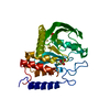
| ||||||||
|---|---|---|---|---|---|---|---|---|---|
| 1 |
| ||||||||
| Unit cell |
|
-Components
| #1: Protein | / YOP51 / YOP2B / PASTEURELLA X / PTPASE / YOP51DELTA162 Mass: 33553.883 Da / Num. of mol.: 1 / Fragment: CATALYTIC DOMAIN, RESIDUES 163 - 468 Source method: isolated from a genetically manipulated source Details: TUNGSTATE AND SULFATE LIGANDS / Source: (gene. exp.) Yersinia enterocolitica (bacteria) / Strain: W22703 / Cell line: BL21 / Gene: YOP51 / Plasmid: PT7-7 / Species (production host): Escherichia coli / Gene (production host): YOP51 / Production host: Escherichia coli BL21(DE3) (bacteria) / Strain (production host): BL21 (DE3) / References: UniProt: P15273, protein-tyrosine-phosphatase |
|---|---|
| #2: Chemical | ChemComp-WO4 /   Mass: 247.838 Da / Num. of mol.: 1 / Source method: obtained synthetically / Formula: WO4 Mass: 247.838 Da / Num. of mol.: 1 / Source method: obtained synthetically / Formula: WO4 |
| #3: Chemical | ChemComp-SO4 / Sulfate  Mass: 96.063 Da / Num. of mol.: 1 / Source method: obtained synthetically / Formula: SO4 Mass: 96.063 Da / Num. of mol.: 1 / Source method: obtained synthetically / Formula: SO4 |
| #4: Water | ChemComp-HOH / Water Mass: 18.015 Da / Num. of mol.: 139 / Source method: isolated from a natural source / Formula: H2O Mass: 18.015 Da / Num. of mol.: 139 / Source method: isolated from a natural source / Formula: H2O |
-Experimental details
-Experiment
| Experiment | Method: X-RAY DIFFRACTION / Number of used crystals: 4 |
|---|
- Sample preparation
| Crystal | Density Matthews: 2.1 Å3/Da / Density % sol: 52 % | |||||||||||||||||||||||||||||||||||||||||||||||||||||||||||||||
|---|---|---|---|---|---|---|---|---|---|---|---|---|---|---|---|---|---|---|---|---|---|---|---|---|---|---|---|---|---|---|---|---|---|---|---|---|---|---|---|---|---|---|---|---|---|---|---|---|---|---|---|---|---|---|---|---|---|---|---|---|---|---|---|---|
| Crystal grow | Temperature: 296 K / Method: vapor diffusion, hanging drop / pH: 8.5 Details: THE CATALYTIC DOMAIN (RESIDUES 163 - 468) OF YOP51 WAS CRYSTALLIZED AT 20 MG/ML WITH 1 MM SODIUM TUNGSTATE AT 23 DEGREES CELSIUS, BY HANGING DROP VAPOR DIFFUSION AGAINST A WELL SOLUTION OF ...Details: THE CATALYTIC DOMAIN (RESIDUES 163 - 468) OF YOP51 WAS CRYSTALLIZED AT 20 MG/ML WITH 1 MM SODIUM TUNGSTATE AT 23 DEGREES CELSIUS, BY HANGING DROP VAPOR DIFFUSION AGAINST A WELL SOLUTION OF 22% POLYETHYLENE GLYCOL (MW 4000), 200 MM LITHIUM SULFATE, 10% ISOPROPANOL, 100 MM TRIS HCL (PH 8.5) AND 0.1% BETA-MERCAPTOETHANOL., vapor diffusion - hanging drop, temperature 296K | |||||||||||||||||||||||||||||||||||||||||||||||||||||||||||||||
| Crystal grow | *PLUS Method: vapor diffusion | |||||||||||||||||||||||||||||||||||||||||||||||||||||||||||||||
| Components of the solutions | *PLUS
|
-Data collection
| Diffraction source | Wavelength: 1.5418 |
|---|---|
| Detector | Type: XUONG-HAMLIN MULTIWIRE / Detector: AREA DETECTOR |
| Radiation | Monochromatic (M) / Laue (L): M / Scattering type: x-ray |
| Radiation wavelength | Wavelength: 1.5418 Å / Relative weight: 1 |
| Reflection | Num. obs: 9875 / % possible obs: 89 % / Observed criterion σ(I): 0 / Redundancy: 4 % / Rmerge(I) obs: 0.107 |
| Reflection | *PLUS Highest resolution: 2.4 Å / Lowest resolution: 9999 Å |
| Reflection shell | *PLUS Highest resolution: 2.4 Å / Lowest resolution: 2.5 Å / Rmerge(I) obs: 0.274 |
- Processing
| Software |
| ||||||||||||||||||||||||||||||||||||||||||||||||||||||||||||||||||||||||||||||||
|---|---|---|---|---|---|---|---|---|---|---|---|---|---|---|---|---|---|---|---|---|---|---|---|---|---|---|---|---|---|---|---|---|---|---|---|---|---|---|---|---|---|---|---|---|---|---|---|---|---|---|---|---|---|---|---|---|---|---|---|---|---|---|---|---|---|---|---|---|---|---|---|---|---|---|---|---|---|---|---|---|---|
| Refinement | Method to determine structure: MIR / Resolution: 2.4→7 Å / σ(F): 0
| ||||||||||||||||||||||||||||||||||||||||||||||||||||||||||||||||||||||||||||||||
| Displacement parameters | Biso mean: 16 Å2
| ||||||||||||||||||||||||||||||||||||||||||||||||||||||||||||||||||||||||||||||||
| Refinement step | Cycle: LAST / Resolution: 2.4→7 Å
| ||||||||||||||||||||||||||||||||||||||||||||||||||||||||||||||||||||||||||||||||
| Refine LS restraints |
| ||||||||||||||||||||||||||||||||||||||||||||||||||||||||||||||||||||||||||||||||
| LS refinement shell | Resolution: 2.4→2.5 Å /
| ||||||||||||||||||||||||||||||||||||||||||||||||||||||||||||||||||||||||||||||||
| Xplor file |
| ||||||||||||||||||||||||||||||||||||||||||||||||||||||||||||||||||||||||||||||||
| Software | *PLUS Name: X-PLOR / Classification: refinement | ||||||||||||||||||||||||||||||||||||||||||||||||||||||||||||||||||||||||||||||||
| Refinement | *PLUS | ||||||||||||||||||||||||||||||||||||||||||||||||||||||||||||||||||||||||||||||||
| Solvent computation | *PLUS | ||||||||||||||||||||||||||||||||||||||||||||||||||||||||||||||||||||||||||||||||
| Displacement parameters | *PLUS | ||||||||||||||||||||||||||||||||||||||||||||||||||||||||||||||||||||||||||||||||
| Refine LS restraints | *PLUS
|
