 Movie
Movie Controller

[English] 日本語
 Yorodumi
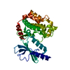
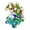
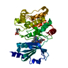
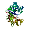
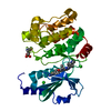
- PDB-1yhw: Crystal Structure of PAK1 kinase domain with one point mutations ... -
+ Open data

- Basic information
| Entry | Database: PDB / ID: 1yhw | ||||||
|---|---|---|---|---|---|---|---|
| Title | Crystal Structure of PAK1 kinase domain with one point mutations (K299R) | ||||||
 Components Components | Serine/threonine-protein kinase PAK 1 | ||||||
 Keywords Keywords |  SIGNALING PROTEIN / TRANSFERASE / kinase / active conformation / activation loop / ATP binding site SIGNALING PROTEIN / TRANSFERASE / kinase / active conformation / activation loop / ATP binding site | ||||||
| Function / homology |  Function and homology information Function and homology informationnegative regulation of cell proliferation involved in contact inhibition / protein localization to cytoplasmic stress granule / positive regulation of microtubule nucleation / hepatocyte growth factor receptor signaling pathway / RHO GTPases Activate ROCKs / gamma-tubulin binding / Activation of RAC1 / CD28 dependent Vav1 pathway / Ephrin signaling / positive regulation of intracellular estrogen receptor signaling pathway ...negative regulation of cell proliferation involved in contact inhibition / protein localization to cytoplasmic stress granule / positive regulation of microtubule nucleation / hepatocyte growth factor receptor signaling pathway / RHO GTPases Activate ROCKs / gamma-tubulin binding / Activation of RAC1 / CD28 dependent Vav1 pathway / Ephrin signaling / positive regulation of intracellular estrogen receptor signaling pathway / DSCAM interactions / RHOV GTPase cycle / regulation of axonogenesis / branching morphogenesis of an epithelial tube / Fc-gamma receptor signaling pathway involved in phagocytosis / RHOJ GTPase cycle / RHOQ GTPase cycle / exocytosis / RHO GTPases activate PAKs / stimulatory C-type lectin receptor signaling pathway / regulation of MAPK cascade / RHOH GTPase cycle / CDC42 GTPase cycle / RHOU GTPase cycle / Sema3A PAK dependent Axon repulsion / Generation of second messenger molecules / intercalated disc / Smooth Muscle Contraction / RAC3 GTPase cycle / RAC2 GTPase cycle / ephrin receptor signaling pathway / localization / positive regulation of microtubule polymerization / RHO GTPases activate PKNs / positive regulation of JUN kinase activity / positive regulation of stress fiber assembly / ruffle / RAC1 GTPase cycle / EPHB-mediated forward signaling / collagen binding / CD209 (DC-SIGN) signaling / neuron projection morphogenesis / VEGFR2 mediated vascular permeability / Signal transduction by L1 / actin filament / regulation of actin cytoskeleton organization / FCERI mediated MAPK activation / MAPK6/MAPK4 signaling / wound healing / G beta:gamma signalling through CDC42 / Regulation of actin dynamics for phagocytic cup formation / ruffle membrane / Z disc / cell-cell junction / cell migration / lamellipodium / positive regulation of peptidyl-serine phosphorylation / chromosome / actin cytoskeleton organization / nuclear membrane / protein autophosphorylation / non-specific serine/threonine protein kinase / protein kinase activity / positive regulation of cell migration / intracellular signal transduction / chromatin remodeling / positive regulation of protein phosphorylation / axon / phosphorylation / protein phosphorylation / focal adhesion / protein serine kinase activity / centrosome / protein serine/threonine kinase activity / dendrite / apoptotic process / DNA damage response / positive regulation of cell population proliferation / protein-containing complex / nucleoplasm / ATP binding / identical protein binding / plasma membrane / cytosol / cytoplasmSimilarity search - Function | ||||||
| Biological species |  Homo sapiens (human) Homo sapiens (human) | ||||||
| Method | X-RAY DIFFRACTION / SYNCHROTRON / MOLECULAR REPLACEMENT / Resolution: 1.8 Å | ||||||
 Authors Authors | Lei, M. / Robinson, M.A. / Harrison, S.C. | ||||||
 Citation Citation | Journal: Structure / Year: 2005 Title: The Active Conformation of the PAK1 Kinase Domain Authors: Lei, M. / Robinson, M.A. / Harrison, S.C. | ||||||
| History |
|
- Structure visualization
| Structure viewer | Molecule: MolmilJmol/JSmol |
|---|
- Downloads & links
-Download
| PDBx/mmCIF format | 1yhw.cif.gz | 76.8 KB | Display | PDBx/mmCIF format |
|---|---|---|---|---|
| PDB format | pdb1yhw.ent.gz | 56.4 KB | Display | PDB format |
| PDBx/mmJSON format | 1yhw.json.gz | Tree view | PDBx/mmJSON format | |
| Others |  Other downloads Other downloads |
-Validation report
| Arichive directory | https://data.pdbj.org/pub/pdb/validation_reports/yh/1yhwftp://data.pdbj.org/pub/pdb/validation_reports/yh/1yhw | HTTPS FTP |
|---|
-Related structure data


-Links
 PDBj
PDBj


- Assembly
| Deposited unit | 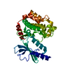
| ||||||||||
|---|---|---|---|---|---|---|---|---|---|---|---|
| 1 |
| ||||||||||
| Unit cell |
|
-Components
| #1: Protein | Mass: 33273.234 Da / Num. of mol.: 1 / Fragment: kinase domain / Mutation: K299R Source method: isolated from a genetically manipulated source Source: (gene. exp.) Homo sapiens (human) / Gene: PAK1 / Plasmid: pGEX2T / Production host:  Escherichia coli (E. coli) / Strain (production host): NB42 / References: UniProt: Q13153, EC: 2.7.1.37 Escherichia coli (E. coli) / Strain (production host): NB42 / References: UniProt: Q13153, EC: 2.7.1.37 |
|---|---|
| #2: Water | ChemComp-HOH / Water Mass: 18.015 Da / Num. of mol.: 349 / Source method: isolated from a natural source / Formula: H2O Mass: 18.015 Da / Num. of mol.: 349 / Source method: isolated from a natural source / Formula: H2O |
-Experimental details
-Experiment
| Experiment | Method: X-RAY DIFFRACTION / Number of used crystals: 1 |
|---|
- Sample preparation
| Crystal | Density Matthews: 2.5 Å3/Da / Density % sol: 50.4 % |
|---|---|
| Crystal grow | Temperature: 277 K / Method: vapor diffusion, hanging drop / pH: 6.5 Details: PEG 4000, NaCl, PIPES, pH 6.5, VAPOR DIFFUSION, HANGING DROP, temperature 277K |
-Data collection
| Diffraction | Mean temperature: 113 K |
|---|---|
| Diffraction source | Source: SYNCHROTRON / Site: APS  / Beamline: 14-BM-C / Wavelength: 1 Å / Beamline: 14-BM-C / Wavelength: 1 Å |
| Detector | Type: ADSC QUANTUM 4 / Detector: CCD / Date: Jan 1, 2000 |
| Radiation | Protocol: SINGLE WAVELENGTH / Monochromatic (M) / Laue (L): M / Scattering type: x-ray |
| Radiation wavelength | Wavelength: 1 Å / Relative weight: 1 |
| Reflection | Resolution: 1.8→50 Å / Num. all: 29341 / Num. obs: 28485 / % possible obs: 92.4 % / Observed criterion σ(I): -3 / Redundancy: 4.3 % / Rmerge(I) obs: 0.073 / Rsym value: 0.344 / Net I/σ(I): 16.9 |
- Processing
| Software |
| ||||||||||||||||||||||||||||
|---|---|---|---|---|---|---|---|---|---|---|---|---|---|---|---|---|---|---|---|---|---|---|---|---|---|---|---|---|---|
| Refinement | Method to determine structure: MOLECULAR REPLACEMENT / Resolution: 1.8→50 Å / Data cutoff high absF: 1000 / Data cutoff low absF: 0 / Cross valid method: THROUGHOUT / σ(F): 0 / Stereochemistry target values: Engh & Huber
| ||||||||||||||||||||||||||||
| Displacement parameters |
| ||||||||||||||||||||||||||||
| Refinement step | Cycle: LAST / Resolution: 1.8→50 Å
| ||||||||||||||||||||||||||||
| Refine LS restraints |
|
