 Movie
Movie Controller


+ Open data

- Basic information
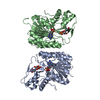
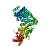
| Entry | Database: PDB / ID: 1ygr | ||||||
|---|---|---|---|---|---|---|---|
| Title | Crystal structure of the tandem phosphatase domain of RPTP CD45 | ||||||
 Components Components |
| ||||||
 Keywords Keywords |  HYDROLASE / Protein tyrosine Phosphatase / RPTP / CD45 / LCA / lymphocyte activation / CD3 zeta / ITAM HYDROLASE / Protein tyrosine Phosphatase / RPTP / CD45 / LCA / lymphocyte activation / CD3 zeta / ITAM | ||||||
| Function / homology |  Function and homology information Function and homology informationplasma membrane raft distribution / positive regulation of antigen receptor-mediated signaling pathway / membrane microdomain / regulation of protein tyrosine kinase activity / positive regulation of protein tyrosine phosphatase activity / positive regulation of hematopoietic stem cell migration / negative regulation of cytokine-mediated signaling pathway / alpha-beta T cell proliferation / negative regulation of protein tyrosine kinase activity / positive regulation of Fc receptor mediated stimulatory signaling pathway ...plasma membrane raft distribution / positive regulation of antigen receptor-mediated signaling pathway / membrane microdomain / regulation of protein tyrosine kinase activity / positive regulation of protein tyrosine phosphatase activity / positive regulation of hematopoietic stem cell migration / negative regulation of cytokine-mediated signaling pathway / alpha-beta T cell proliferation / negative regulation of protein tyrosine kinase activity / positive regulation of Fc receptor mediated stimulatory signaling pathway / negative regulation of cell adhesion involved in substrate-bound cell migration / gamma-delta T cell receptor complex / regulation of interleukin-8 production / Fc-gamma receptor III complex / negative regulation of microglial cell activation / Other semaphorin interactions / negative regulation of T cell mediated cytotoxicity / positive regulation of humoral immune response mediated by circulating immunoglobulin / DN2 thymocyte differentiation / cell cycle phase transition / negative regulation of protein autophosphorylation / gamma-delta T cell differentiation / positive regulation of gamma-delta T cell differentiation / natural killer cell differentiation / transmembrane receptor protein tyrosine phosphatase activity / Fc-gamma receptor signaling pathway / cellular response to extracellular stimulus / bleb / gamma-delta T cell activation / positive regulation of alpha-beta T cell proliferation / positive regulation of isotype switching to IgG isotypes / negative thymic T cell selection / stem cell development / heparan sulfate proteoglycan binding / positive regulation of protein localization to cell surface / alpha-beta T cell receptor complex / positive thymic T cell selection / regulation of phagocytosis / Nef and signal transduction / positive regulation of extrinsic apoptotic signaling pathway / heterotypic cell-cell adhesion / bone marrow development / regulation of receptor signaling pathway via JAK-STAT / T cell receptor complex / negative regulation of interleukin-2 production / ankyrin binding / leukocyte cell-cell adhesion / spectrin binding / response to aldosterone / positive regulation of stem cell proliferation / Translocation of ZAP-70 to Immunological synapse / Phosphorylation of CD3 and TCR zeta chains / B cell proliferation / : / alpha-beta T cell activation / positive regulation of immunoglobulin production / Generation of second messenger molecules / FCGR activation / T cell differentiation / PD-1 signaling / positive regulation of protein kinase activity / Role of phospholipids in phagocytosis / hematopoietic progenitor cell differentiation / positive regulation of phagocytosis / dephosphorylation / positive regulation of B cell proliferation / extrinsic apoptotic signaling pathway / release of sequestered calcium ion into cytosol / positive regulation of T cell proliferation / protein tyrosine kinase binding / positive regulation of interleukin-2 production / T cell activation / protein dephosphorylation / FCGR3A-mediated IL10 synthesis / B cell differentiation / protein-tyrosine-phosphatase / secretory granule membrane / protein tyrosine phosphatase activity / response to gamma radiation / FCGR3A-mediated phagocytosis / B cell receptor signaling pathway / negative regulation of protein kinase activity / cytoplasmic side of plasma membrane / negative regulation of ERK1 and ERK2 cascade / Regulation of actin dynamics for phagocytic cup formation / positive regulation of T cell mediated cytotoxicity / Immunoregulatory interactions between a Lymphoid and a non-Lymphoid cell / MAPK cascade / positive regulation of peptidyl-tyrosine phosphorylation / transmembrane signaling receptor activity / positive regulation of tumor necrosis factor production / Downstream TCR signaling / protein complex oligomerization / heparin binding / T cell receptor signaling pathway / regulation of gene expression / protein-containing complex assembly / defense response to virus / positive regulation of MAPK cascade / adaptive immune responseSimilarity search - Function | ||||||
| Biological species |  Homo sapiens (human) Homo sapiens (human) | ||||||
| Method | X-RAY DIFFRACTION / SYNCHROTRON / MOLECULAR REPLACEMENT / Resolution: 2.9 Å | ||||||
 Authors Authors | Nam, H.J. / Poy, F. / Saito, H. / Frederick, C.A. | ||||||
 Citation Citation | Journal: J.Exp.Med. / Year: 2005 Title: Structural basis for the function and regulation of the receptor protein tyrosine phosphatase CD45. Authors: Nam, H.J. / Poy, F. / Saito, H. / Frederick, C.A. | ||||||
| History |
|
- Structure visualization
| Structure viewer | Molecule: MolmilJmol/JSmol |
|---|
- Downloads & links
-Download
| PDBx/mmCIF format | 1ygr.cif.gz | 240.5 KB | Display | PDBx/mmCIF format |
|---|---|---|---|---|
| PDB format | pdb1ygr.ent.gz | 199.9 KB | Display | PDB format |
| PDBx/mmJSON format | 1ygr.json.gz | Tree view | PDBx/mmJSON format | |
| Others |  Other downloads Other downloads |
-Validation report
| Arichive directory | https://data.pdbj.org/pub/pdb/validation_reports/yg/1ygrftp://data.pdbj.org/pub/pdb/validation_reports/yg/1ygr | HTTPS FTP |
|---|
-Related structure data


-Links
 PDBj
PDBj


- Assembly
| Deposited unit | 
| ||||||||
|---|---|---|---|---|---|---|---|---|---|
| 1 | 
| ||||||||
| 2 | 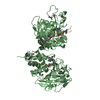
| ||||||||
| Unit cell |
|
-Components
| #1: Protein | PTPRC Mass: 71500.625 Da / Num. of mol.: 2 / Fragment: Cytoplasmic domain / Mutation: C828S Source method: isolated from a genetically manipulated source Source: (gene. exp.) Homo sapiens (human) / Plasmid: pET11 / Species (production host): Escherichia coli / Production host:  Escherichia coli BL21(DE3) (bacteria) / Strain (production host): BL21(DE3) / References: UniProt: P08575 Escherichia coli BL21(DE3) (bacteria) / Strain (production host): BL21(DE3) / References: UniProt: P08575#2: Protein/peptide | Mass: 890.808 Da / Num. of mol.: 2 / Fragment: ITAM-1 (residues 80-85; SWS:P20963) / Source method: obtained synthetically / Details: This sequence occurs naturally in humans. / References: UniProt: P20963 |
|---|
-Experimental details
-Experiment
| Experiment | Method: X-RAY DIFFRACTION |
|---|
- Sample preparation
| Crystal | Density Matthews: 2.81 Å3/Da / Density % sol: 56.19 % |
|---|---|
| Crystal grow | Temperature: 293 K / Method: vapor diffusion, hanging drop / pH: 5 Details: Sodium acetate, PEG 4000, Glycerol, pH 5.0, VAPOR DIFFUSION, HANGING DROP, temperature 293K |
-Data collection
| Diffraction | Mean temperature: 200 K |
|---|---|
| Diffraction source | Source: SYNCHROTRON / Site: NSLS  / Beamline: X12C / Wavelength: 1.0332 Å / Beamline: X12C / Wavelength: 1.0332 Å |
| Detector | Type: ENRAF-NONIUS FAST / Detector: DIFFRACTOMETER / Date: Dec 15, 2002 |
| Radiation | Protocol: SINGLE WAVELENGTH / Monochromatic (M) / Laue (L): M / Scattering type: x-ray |
| Radiation wavelength | Wavelength: 1.0332 Å / Relative weight: 1 |
| Reflection | Resolution: 2.9→29.85 Å / Num. obs: 36165 / Biso Wilson estimate: 59.5 Å2 |
- Processing
| Software |
| |||||||||||||||||||||||||
|---|---|---|---|---|---|---|---|---|---|---|---|---|---|---|---|---|---|---|---|---|---|---|---|---|---|---|
| Refinement | Method to determine structure: MOLECULAR REPLACEMENT / Resolution: 2.9→29.85 Å / Rfactor Rfree error: 0.008 / Data cutoff high absF: 2020965.81 / Data cutoff low absF: 0 / Isotropic thermal model: RESTRAINED / Cross valid method: THROUGHOUT / σ(F): 0 / Stereochemistry target values: Engh & Huber
| |||||||||||||||||||||||||
| Solvent computation | Solvent model: FLAT MODEL / Bsol: 20.7118 Å2 / ksol: 0.26757 e/Å3 | |||||||||||||||||||||||||
| Displacement parameters | Biso mean: 83.2 Å2
| |||||||||||||||||||||||||
| Refine analyze |
| |||||||||||||||||||||||||
| Refinement step | Cycle: LAST / Resolution: 2.9→29.85 Å
| |||||||||||||||||||||||||
| Refine LS restraints |
| |||||||||||||||||||||||||
| Refine LS restraints NCS | NCS model details: CONSTR | |||||||||||||||||||||||||
| LS refinement shell | Resolution: 2.9→3.08 Å / Rfactor Rfree error: 0.045 / Total num. of bins used: 6
| |||||||||||||||||||||||||
| Xplor file |
|
