 Movie
Movie Controller


+ Open data

- Basic information
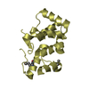
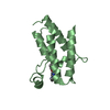
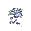
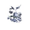
| Entry | Database: PDB / ID: 1y2g | ||||||
|---|---|---|---|---|---|---|---|
| Title | Crystal STructure of ZipA in complex with an inhibitor | ||||||
 Components Components | Cell division protein zipA | ||||||
 Keywords Keywords | CELL CYCLE | ||||||
| Function / homology |  Function and homology informationdivisome complex / FtsZ-dependent cytokinesis / division septum assembly / cell division site / cell division / protein homodimerization activity / plasma membrane Function and homology informationdivisome complex / FtsZ-dependent cytokinesis / division septum assembly / cell division site / cell division / protein homodimerization activity / plasma membraneSimilarity search - Function | ||||||
| Biological species |  Escherichia coli (E. coli) Escherichia coli (E. coli) | ||||||
| Method | X-RAY DIFFRACTION / MOLECULAR REPLACEMENT / Resolution: 1.9 Å | ||||||
 Authors Authors | Mosyak, L. / Rush, T.S. | ||||||
 Citation Citation | Journal: J.Med.Chem. / Year: 2005 Title: A shape-based 3-D scaffold hopping method and its application to a bacterial protein-protein interaction Authors: Rush, T.S. / Grant, J.A. / Mosyak, L. / Nicholls, A. | ||||||
| History |
|
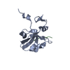
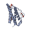
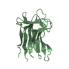
- Structure visualization
| Structure viewer | Molecule: MolmilJmol/JSmol |
|---|
- Downloads & links
-Download
| PDBx/mmCIF format | 1y2g.cif.gz | 71.2 KB | Display | PDBx/mmCIF format |
|---|---|---|---|---|
| PDB format | pdb1y2g.ent.gz | 52.6 KB | Display | PDB format |
| PDBx/mmJSON format | 1y2g.json.gz | Tree view | PDBx/mmJSON format | |
| Others |  Other downloads Other downloads |
-Validation report
| Arichive directory | https://data.pdbj.org/pub/pdb/validation_reports/y2/1y2gftp://data.pdbj.org/pub/pdb/validation_reports/y2/1y2g | HTTPS FTP |
|---|
-Related structure data
| Related structure data |  1y2fC  1f46S C: citing same article ( S: Starting model for refinement |
|---|---|
| Similar structure data |


-Links
 PDBj
PDBj- Assembly
| Deposited unit | 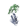
| ||||||||
|---|---|---|---|---|---|---|---|---|---|
| 1 | 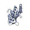
| ||||||||
| 2 | 
| ||||||||
| Unit cell |
|
-Components
| #1: Protein | Mass: 15662.851 Da / Num. of mol.: 2 Source method: isolated from a genetically manipulated source Source: (gene. exp.) Escherichia coli (E. coli) / Gene: zipa / References: UniProt: P77173#2: Chemical | ChemComp-CL3 / | 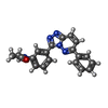  Mass: 343.382 Da / Num. of mol.: 1 / Source method: obtained synthetically / Formula: C20H17N5O Mass: 343.382 Da / Num. of mol.: 1 / Source method: obtained synthetically / Formula: C20H17N5O#3: Water | ChemComp-HOH / | Water Mass: 18.015 Da / Num. of mol.: 223 / Source method: isolated from a natural source / Formula: H2O Mass: 18.015 Da / Num. of mol.: 223 / Source method: isolated from a natural source / Formula: H2O |
|---|
-Experimental details
-Experiment
| Experiment | Method: X-RAY DIFFRACTION |
|---|
- Sample preparation
| Crystal | Density Matthews: 2.26 Å3/Da / Density % sol: 45.49 % |
|---|---|
| Crystal grow | Temperature: 298 K / Method: vapor diffusion / pH: 6 Details: PEG 6000, MES, pH 6.0, VAPOR DIFFUSION, temperature 298.0K |
-Data collection
| Radiation | Protocol: SINGLE WAVELENGTH / Monochromatic (M) / Laue (L): M / Scattering type: x-ray |
|---|---|
| Radiation wavelength | Relative weight: 1 |
| Reflection | Resolution: 1.9→25 Å / Num. obs: 22159 / Biso Wilson estimate: 28.2 Å2 |
| Reflection shell | Resolution: 1.9→1.97 Å |
- Processing
| Software | Name: CNS / Version: 1.1 / Classification: refinement | ||||||||||||||||||||||||||||||||||||||||||||||||||||||||||||||||||||||||||||||||
|---|---|---|---|---|---|---|---|---|---|---|---|---|---|---|---|---|---|---|---|---|---|---|---|---|---|---|---|---|---|---|---|---|---|---|---|---|---|---|---|---|---|---|---|---|---|---|---|---|---|---|---|---|---|---|---|---|---|---|---|---|---|---|---|---|---|---|---|---|---|---|---|---|---|---|---|---|---|---|---|---|---|
| Refinement | Method to determine structure: MOLECULAR REPLACEMENT Starting model: PDB ENTRY 1F46 Resolution: 1.9→23.48 Å / Rfactor Rfree error: 0.007 / Data cutoff high absF: 606413.33 / Data cutoff low absF: 0 / Isotropic thermal model: RESTRAINED / Cross valid method: THROUGHOUT / σ(F): 0 / Stereochemistry target values: Engh & Huber
| ||||||||||||||||||||||||||||||||||||||||||||||||||||||||||||||||||||||||||||||||
| Solvent computation | Solvent model: FLAT MODEL / Bsol: 48.2251 Å2 / ksol: 0.338309 e/Å3 | ||||||||||||||||||||||||||||||||||||||||||||||||||||||||||||||||||||||||||||||||
| Displacement parameters | Biso mean: 39.7 Å2
| ||||||||||||||||||||||||||||||||||||||||||||||||||||||||||||||||||||||||||||||||
| Refine analyze |
| ||||||||||||||||||||||||||||||||||||||||||||||||||||||||||||||||||||||||||||||||
| Refinement step | Cycle: LAST / Resolution: 1.9→23.48 Å
| ||||||||||||||||||||||||||||||||||||||||||||||||||||||||||||||||||||||||||||||||
| Refine LS restraints |
| ||||||||||||||||||||||||||||||||||||||||||||||||||||||||||||||||||||||||||||||||
| LS refinement shell | Resolution: 1.9→2.02 Å / Rfactor Rfree error: 0.028 / Total num. of bins used: 6
| ||||||||||||||||||||||||||||||||||||||||||||||||||||||||||||||||||||||||||||||||
| Xplor file |
|
