 Movie
Movie Controller


+ Open data

- Basic information
| Entry | Database: PDB / ID: 1xtc | ||||||
|---|---|---|---|---|---|---|---|
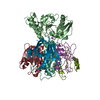
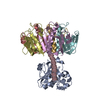
| Title | CHOLERA TOXIN | ||||||
 Components Components | (CHOLERA TOXIN ) x 3 ) x 3 | ||||||
 Keywords Keywords | TOXIN / ENTEROTOXIN | ||||||
| Function / homology |  Function and homology information Function and homology informationhost cell surface binding / galactose binding / glycosyltransferase activity / Transferases; Glycosyltransferases; Pentosyltransferases / catalytic complex / localization / positive regulation of tyrosine phosphorylation of STAT protein / nucleotidyltransferase activity / toxin activity / periplasmic space ...host cell surface binding / galactose binding / glycosyltransferase activity / Transferases; Glycosyltransferases; Pentosyltransferases / catalytic complex / localization / positive regulation of tyrosine phosphorylation of STAT protein / nucleotidyltransferase activity / toxin activity / periplasmic space / lipid binding / host cell plasma membrane / extracellular space / extracellular region / membraneSimilarity search - Function | ||||||
| Biological species |   Vibrio cholerae (bacteria) Vibrio cholerae (bacteria) | ||||||
| Method | X-RAY DIFFRACTION / Resolution: 2.4 Å | ||||||
 Authors Authors | Zhang, R.-G. / Westbrook, E. | ||||||
 Citation Citation | Journal: J.Mol.Biol. / Year: 1995 Title: The three-dimensional crystal structure of cholera toxin. Authors: Zhang, R.G. / Scott, D.L. / Westbrook, M.L. / Nance, S. / Spangler, B.D. / Shipley, G.G. / Westbrook, E.M. #1: Journal: J.Mol.Biol. / Year: 1995Title: The 2.4 A Crystal Structure of Cholera Toxin B Subunit Pentamer: Choleragenoid Authors: Zhang, R.G. / Westbrook, M.L. / Westbrook, E.M. / Scott, D.L. / Otwinowski, Z. / Maulik, P.R. / Reed, R.A. / Shipley, G.G. | ||||||
| History |
|
- Structure visualization
| Structure viewer | Molecule: MolmilJmol/JSmol |
|---|
- Downloads & links
-Download
| PDBx/mmCIF format | 1xtc.cif.gz | 156.8 KB | Display | PDBx/mmCIF format |
|---|---|---|---|---|
| PDB format | pdb1xtc.ent.gz | 129.9 KB | Display | PDB format |
| PDBx/mmJSON format | 1xtc.json.gz | Tree view | PDBx/mmJSON format | |
| Others |  Other downloads Other downloads |
-Validation report
| Arichive directory | https://data.pdbj.org/pub/pdb/validation_reports/xt/1xtcftp://data.pdbj.org/pub/pdb/validation_reports/xt/1xtc | HTTPS FTP |
|---|
-Related structure data
| Similar structure data |
|---|


-Links
 PDBj
PDBj
- Assembly
| Deposited unit | 
| ||||||||
|---|---|---|---|---|---|---|---|---|---|
| 1 |
| ||||||||
| Unit cell |
|
-Components
| #1: Protein | / CTX / CHOLERAGEN Mass: 21841.975 Da / Num. of mol.: 1 / Source method: isolated from a natural source Details: COMMERCIALLY OBTAINED FROM LIST BIOLOGICAL LABORATORY, CAMPBER CA95008 Source: (natural) Vibrio cholerae (bacteria) / Strain: 569B / References: UniProt: P01555 | ||||
|---|---|---|---|---|---|
| #2: Protein/peptide | / CTX / CHOLERAGEN Mass: 5404.996 Da / Num. of mol.: 1 / Source method: isolated from a natural source Details: COMMERCIALLY OBTAINED FROM LIST BIOLOGICAL LABORATORY, CAMPBER CA95008 Source: (natural) Vibrio cholerae (bacteria) / Strain: 569B / References: UniProt: P01555 | ||||
| #3: Protein | / CTX / CHOLERAGEN Mass: 11692.346 Da / Num. of mol.: 5 / Source method: isolated from a natural source Details: COMMERCIALLY OBTAINED FROM LIST BIOLOGICAL LABORATORY, CAMPBER CA95008 Source: (natural) Vibrio cholerae (bacteria) / Strain: 569B / References: UniProt: P01556#4: Water | ChemComp-HOH / | Water Mass: 18.015 Da / Num. of mol.: 138 / Source method: isolated from a natural source / Formula: H2O Mass: 18.015 Da / Num. of mol.: 138 / Source method: isolated from a natural source / Formula: H2OCompound details | CHOLERA TOXIN CONTAINS 3 KINDS OF CHAINS: AN ALPHA AND A GAMMA CHAIN (FROM THE SAME PRECURSOR ...CHOLERA TOXIN CONTAINS 3 KINDS OF CHAINS: AN ALPHA AND A GAMMA CHAIN (FROM THE SAME PRECURSOR MOLECULE), LINKED BY AN INTERCHAIN | |
-Experimental details
-Experiment
| Experiment | Method: X-RAY DIFFRACTION |
|---|
- Sample preparation
| Crystal | Density Matthews: 2.28 Å3/Da / Density % sol: 46.1 % |
|---|---|
| Crystal grow | *PLUS Method: other / Details: Spangler, B.D., (1989) Biochemistry, 28, 1333. |
-Data collection
| Diffraction source | Wavelength: 1.5418 |
|---|---|
| Detector | Type: SIEMENS-NICOLET X100 / Detector: AREA DETECTOR / Date: Aug 15, 1989 |
| Radiation | Monochromatic (M) / Laue (L): M / Scattering type: x-ray |
| Radiation wavelength | Wavelength: 1.5418 Å / Relative weight: 1 |
| Reflection | Num. obs: 26200 / % possible obs: 88.4 % / Rmerge(I) obs: 0.048 |
| Reflection | *PLUS Highest resolution: 2.4 Å / Num. measured all: 60810 / Rmerge(I) obs: 0.048 |
- Processing
| Software |
| ||||||||||||||||||||||||||||||||||||||||||||||||||||||||||||||||||||||||||||||||||||
|---|---|---|---|---|---|---|---|---|---|---|---|---|---|---|---|---|---|---|---|---|---|---|---|---|---|---|---|---|---|---|---|---|---|---|---|---|---|---|---|---|---|---|---|---|---|---|---|---|---|---|---|---|---|---|---|---|---|---|---|---|---|---|---|---|---|---|---|---|---|---|---|---|---|---|---|---|---|---|---|---|---|---|---|---|---|
| Refinement | Highest resolution: 2.4 Å / σ(F): 1.5 /
| ||||||||||||||||||||||||||||||||||||||||||||||||||||||||||||||||||||||||||||||||||||
| Displacement parameters | Biso mean: 20.37 Å2 | ||||||||||||||||||||||||||||||||||||||||||||||||||||||||||||||||||||||||||||||||||||
| Refinement step | Cycle: LAST / Highest resolution: 2.4 Å
| ||||||||||||||||||||||||||||||||||||||||||||||||||||||||||||||||||||||||||||||||||||
| Refine LS restraints |
| ||||||||||||||||||||||||||||||||||||||||||||||||||||||||||||||||||||||||||||||||||||
| Software | *PLUS Name: PROFFT / Classification: refinement | ||||||||||||||||||||||||||||||||||||||||||||||||||||||||||||||||||||||||||||||||||||
| Refinement | *PLUS Lowest resolution: 10 Å / Rfactor obs: 0.185 | ||||||||||||||||||||||||||||||||||||||||||||||||||||||||||||||||||||||||||||||||||||
| Solvent computation | *PLUS | ||||||||||||||||||||||||||||||||||||||||||||||||||||||||||||||||||||||||||||||||||||
| Displacement parameters | *PLUS |
