 Movie
Movie Controller

[English] 日本語
 Yorodumi
- PDB-1xjn: Structural mechanism of allosteric substrate specificity in a rib... -
+ Open data

- Basic information
| Entry | Database: PDB / ID: 1xjn | ||||||
|---|---|---|---|---|---|---|---|
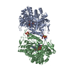
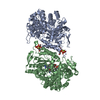
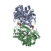
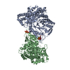
| Title | Structural mechanism of allosteric substrate specificity in a ribonucleotide reductase: dATP-CDP complex | ||||||
 Components Components | ribonucleotide reductase, B12-dependent | ||||||
 Keywords Keywords | OXIDOREDUCTASE / ribonucleotide reductase / 10 alpha-beta barrel / allosteric regulation / substrate specificity / protein-nucleotide complex | ||||||
| Function / homology |  Function and homology informationcobalamin binding / ribonucleoside-diphosphate reductase / ribonucleoside-diphosphate reductase activity, thioredoxin disulfide as acceptor / deoxyribonucleotide biosynthetic process / DNA biosynthetic process / DNA replication / ATP binding Function and homology informationcobalamin binding / ribonucleoside-diphosphate reductase / ribonucleoside-diphosphate reductase activity, thioredoxin disulfide as acceptor / deoxyribonucleotide biosynthetic process / DNA biosynthetic process / DNA replication / ATP bindingSimilarity search - Function | ||||||
| Biological species |   Thermotoga maritima (bacteria) Thermotoga maritima (bacteria) | ||||||
| Method | X-RAY DIFFRACTION / SYNCHROTRON / MOLECULAR REPLACEMENT / Resolution: 2.25 Å | ||||||
 Authors Authors | Larsson, K.-M. / Jordan, A. / Eliasson, R. / Reichard, P. / Logan, D.T. / Nordlund, P. | ||||||
 Citation Citation | Journal: Nat.Struct.Mol.Biol. / Year: 2004 Title: Structural Mechanism of Allosteric Substrate Specificity Regulation in a Ribonucleotide Reductase Authors: Larsson, K.-M. / Jordan, A. / Eliasson, R. / Reichard, P. / Logan, D.T. / Nordlund, P. | ||||||
| History |
| ||||||
| Remark 999 | SEQUENCE Authors suggest a point mutation either in the sequencing clone (SER->TYR mutation) or in ...SEQUENCE Authors suggest a point mutation either in the sequencing clone (SER->TYR mutation) or in their expression clone (TYR->SER). Codons for TYR=ATA and SER=AGA. |
- Structure visualization
| Structure viewer | Molecule: MolmilJmol/JSmol |
|---|
- Downloads & links
-Download
| PDBx/mmCIF format | 1xjn.cif.gz | 505.4 KB | Display | PDBx/mmCIF format |
|---|---|---|---|---|
| PDB format | pdb1xjn.ent.gz | 413.1 KB | Display | PDB format |
| PDBx/mmJSON format | 1xjn.json.gz | Tree view | PDBx/mmJSON format | |
| Others |  Other downloads Other downloads |
-Validation report
| Arichive directory | https://data.pdbj.org/pub/pdb/validation_reports/xj/1xjnftp://data.pdbj.org/pub/pdb/validation_reports/xj/1xjn | HTTPS FTP |
|---|
-Related structure data
| Related structure data |  1xjeSC  1xjfC  1xjgC  1xjjC 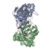 1xjkC  1xjmC S: Starting model for refinement C: citing same article ( |
|---|---|
| Similar structure data |


-Links
 PDBj
PDBj


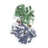
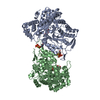
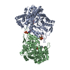
- Assembly
| Deposited unit | 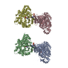
| ||||||||
|---|---|---|---|---|---|---|---|---|---|
| 1 | 
| ||||||||
| 2 | 
| ||||||||
| Unit cell |
| ||||||||
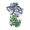
| Details | The asymmetric unit consists of two homodimers. The homodimer constitute is the smallest functional unit. |
-Components
| #1: Protein | Mass: 73374.234 Da / Num. of mol.: 4 / Fragment: residues 1-644 Source method: isolated from a genetically manipulated source Source: (gene. exp.) Thermotoga maritima (bacteria) / Gene: nrdj / Plasmid: pET-22b / Species (production host): Escherichia coli / Production host: Escherichia coli BL21(DE3) (bacteria) / Strain (production host): BL21(DE3)References: UniProt: O33839, ribonucleoside-diphosphate reductase#2: Chemical | ChemComp-CDP / Cytidine diphosphate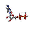  Mass: 403.176 Da / Num. of mol.: 4 / Source method: obtained synthetically / Formula: C9H15N3O11P2 Mass: 403.176 Da / Num. of mol.: 4 / Source method: obtained synthetically / Formula: C9H15N3O11P2#3: Chemical | ChemComp-DTP / Deoxyadenosine triphosphate  Mass: 491.182 Da / Num. of mol.: 4 / Source method: obtained synthetically / Formula: C10H16N5O12P3 Mass: 491.182 Da / Num. of mol.: 4 / Source method: obtained synthetically / Formula: C10H16N5O12P3#4: Chemical | ChemComp-CL / | Chloride  Mass: 35.453 Da / Num. of mol.: 1 / Source method: obtained synthetically / Formula: Cl Mass: 35.453 Da / Num. of mol.: 1 / Source method: obtained synthetically / Formula: Cl#5: Water | ChemComp-HOH / | Water Mass: 18.015 Da / Num. of mol.: 649 / Source method: isolated from a natural source / Formula: H2O Mass: 18.015 Da / Num. of mol.: 649 / Source method: isolated from a natural source / Formula: H2O |
|---|
-Experimental details
-Experiment
| Experiment | Method: X-RAY DIFFRACTION |
|---|
- Sample preparation
| Crystal | Density Matthews: 2.55 Å3/Da / Density % sol: 51.68 % |
|---|---|
| Crystal grow | Temperature: 293 K / Method: vapor diffusion, hanging drop / pH: 4.5 Details: PEG8000, sodium acetate, sodium chloride, dithiotreithol, pH 4.5, VAPOR DIFFUSION, HANGING DROP, temperature 293K |
-Data collection
| Diffraction | Mean temperature: 100 K |
|---|---|
| Diffraction source | Source: SYNCHROTRON / Site: EMBL/DESY, HAMBURG  / Beamline: X11 / Wavelength: 0.8126 Å / Beamline: X11 / Wavelength: 0.8126 Å |
| Detector | Type: MARRESEARCH / Detector: CCD / Date: Jan 24, 2003 |
| Radiation | Protocol: SINGLE WAVELENGTH / Monochromatic (M) / Laue (L): M / Scattering type: x-ray |
| Radiation wavelength | Wavelength: 0.8126 Å / Relative weight: 1 |
| Reflection | Resolution: 2.25→20 Å / Num. all: 139338 / Num. obs: 138333 / % possible obs: 99.3 % / Redundancy: 3 % / Rsym value: 0.097 / Net I/σ(I): 8 |
| Reflection shell | Resolution: 2.25→2.5 Å / Redundancy: 3 % / Mean I/σ(I) obs: 3.2 / Rsym value: 0.368 / % possible all: 99.7 |
- Processing
| Software |
| ||||||||||||||||||||||||||||||||||||||||||||||||||||||||||||||||||||||||||||||||||||||||||||||||||||||||||||||||||||||||||||||||||||||||||||||||||||||||||||||||||||||||||
|---|---|---|---|---|---|---|---|---|---|---|---|---|---|---|---|---|---|---|---|---|---|---|---|---|---|---|---|---|---|---|---|---|---|---|---|---|---|---|---|---|---|---|---|---|---|---|---|---|---|---|---|---|---|---|---|---|---|---|---|---|---|---|---|---|---|---|---|---|---|---|---|---|---|---|---|---|---|---|---|---|---|---|---|---|---|---|---|---|---|---|---|---|---|---|---|---|---|---|---|---|---|---|---|---|---|---|---|---|---|---|---|---|---|---|---|---|---|---|---|---|---|---|---|---|---|---|---|---|---|---|---|---|---|---|---|---|---|---|---|---|---|---|---|---|---|---|---|---|---|---|---|---|---|---|---|---|---|---|---|---|---|---|---|---|---|---|---|---|---|---|---|
| Refinement | Method to determine structure: MOLECULAR REPLACEMENT Starting model: PDB entry 1XJE Resolution: 2.25→19.54 Å / Cor.coef. Fo:Fc: 0.95 / Cor.coef. Fo:Fc free: 0.909 / SU B: 6.822 / SU ML: 0.168 / Cross valid method: THROUGHOUT / ESU R: 0.281 / ESU R Free: 0.23 / Stereochemistry target values: MAXIMUM LIKELIHOOD / Details: HYDROGENS HAVE BEEN ADDED IN THE RIDING POSITIONS
| ||||||||||||||||||||||||||||||||||||||||||||||||||||||||||||||||||||||||||||||||||||||||||||||||||||||||||||||||||||||||||||||||||||||||||||||||||||||||||||||||||||||||||
| Solvent computation | Ion probe radii: 0.8 Å / Shrinkage radii: 0.8 Å / VDW probe radii: 1.2 Å / Solvent model: MASK | ||||||||||||||||||||||||||||||||||||||||||||||||||||||||||||||||||||||||||||||||||||||||||||||||||||||||||||||||||||||||||||||||||||||||||||||||||||||||||||||||||||||||||
| Displacement parameters | Biso mean: 29.101 Å2
| ||||||||||||||||||||||||||||||||||||||||||||||||||||||||||||||||||||||||||||||||||||||||||||||||||||||||||||||||||||||||||||||||||||||||||||||||||||||||||||||||||||||||||
| Refinement step | Cycle: LAST / Resolution: 2.25→19.54 Å
| ||||||||||||||||||||||||||||||||||||||||||||||||||||||||||||||||||||||||||||||||||||||||||||||||||||||||||||||||||||||||||||||||||||||||||||||||||||||||||||||||||||||||||
| Refine LS restraints |
| ||||||||||||||||||||||||||||||||||||||||||||||||||||||||||||||||||||||||||||||||||||||||||||||||||||||||||||||||||||||||||||||||||||||||||||||||||||||||||||||||||||||||||
| LS refinement shell | Resolution: 2.25→2.308 Å / Total num. of bins used: 20
|
