 Movie
Movie Controller

[English] 日本語
 Yorodumi
- PDB-1xgm: METHIONINE AMINOPEPTIDASE FROM HYPERTHERMOPHILE PYROCOCCUS FURIOSUS -
+ Open data

- Basic information
| Entry | Database: PDB / ID: 1xgm | ||||||
|---|---|---|---|---|---|---|---|
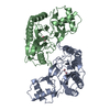
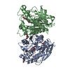
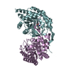
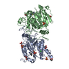
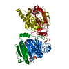
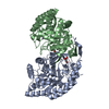
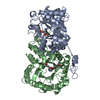
| Title | METHIONINE AMINOPEPTIDASE FROM HYPERTHERMOPHILE PYROCOCCUS FURIOSUS | ||||||
 Components Components | METHIONINE AMINOPEPTIDASE Methionyl aminopeptidase Methionyl aminopeptidase | ||||||
 Keywords Keywords | AMINOPEPTIDASE / HYPERTHERMOPHILE | ||||||
| Function / homology |  Function and homology information Function and homology informationinitiator methionyl aminopeptidase activity / methionyl aminopeptidase / metalloaminopeptidase activity / proteolysis / metal ion bindingSimilarity search - Function | ||||||
| Biological species |   Pyrococcus furiosus (archaea) Pyrococcus furiosus (archaea) | ||||||
| Method | X-RAY DIFFRACTION / SYNCHROTRON / MIR / Resolution: 2.8 Å | ||||||
 Authors Authors | Tahirov, T.H. / Tsukihara, T. | ||||||
 Citation Citation | Journal: J.Mol.Biol. / Year: 1998 Title: Crystal structure of methionine aminopeptidase from hyperthermophile, Pyrococcus furiosus. Authors: Tahirov, T.H. / Oki, H. / Tsukihara, T. / Ogasahara, K. / Yutani, K. / Ogata, K. / Izu, Y. / Tsunasawa, S. / Kato, I. #1: Journal: Acta Crystallogr.,Sect.D / Year: 1997Title: Crystallization and Preliminary X-Ray Analysis of Methionine Aminopeptidase from the Hyperthermophilic Bacterium Pyrococcus Furiosus Authors: Tahirov, T.H. / Oki, H. / Tsukihara, T. / Ogasahara, K. / Izu, Y. / Tsunasawa, S. / Kato, I. / Yutani, K. | ||||||
| History |
|
- Structure visualization
| Structure viewer | Molecule: MolmilJmol/JSmol |
|---|
- Downloads & links
-Download
| PDBx/mmCIF format | 1xgm.cif.gz | 125.1 KB | Display | PDBx/mmCIF format |
|---|---|---|---|---|
| PDB format | pdb1xgm.ent.gz | 98.6 KB | Display | PDB format |
| PDBx/mmJSON format | 1xgm.json.gz | Tree view | PDBx/mmJSON format | |
| Others |  Other downloads Other downloads |
-Validation report
| Arichive directory | https://data.pdbj.org/pub/pdb/validation_reports/xg/1xgmftp://data.pdbj.org/pub/pdb/validation_reports/xg/1xgm | HTTPS FTP |
|---|
-Related structure data


-Links
 PDBj
PDBj
- Assembly
| Deposited unit | 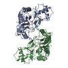
| ||||||||
|---|---|---|---|---|---|---|---|---|---|
| 1 |
| ||||||||
| Unit cell |
| ||||||||
| Noncrystallographic symmetry (NCS) | NCS oper: (Code: given Matrix: (-0.957964, -0.264121, 0.112007), Vector : |
-Components
| #1: Protein | Methionyl aminopeptidase Mass: 32888.383 Da / Num. of mol.: 2 / Source method: isolated from a natural source / Details: TWO COBALT IONS IN ACTIVE SITE / Source: (natural) Pyrococcus furiosus (archaea) / References: UniProt: P56218, methionyl aminopeptidase#2: Chemical | ChemComp-CO /   Mass: 58.933 Da / Num. of mol.: 4 / Source method: obtained synthetically / Formula: Co Mass: 58.933 Da / Num. of mol.: 4 / Source method: obtained synthetically / Formula: Co#3: Water | ChemComp-HOH / | Water Mass: 18.015 Da / Num. of mol.: 10 / Source method: isolated from a natural source / Formula: H2O Mass: 18.015 Da / Num. of mol.: 10 / Source method: isolated from a natural source / Formula: H2O |
|---|
-Experimental details
-Experiment
| Experiment | Method: X-RAY DIFFRACTION / Number of used crystals: 1 |
|---|
- Sample preparation
| Crystal | Density Matthews: 2.39 Å3/Da / Density % sol: 48.8 % Description: THREE HEAVY-ATOM DERIVATIVES USED FOR STRUCTURE DETERMINATION WERE OBTAINED BY SOAKING OF NATIVE CRYSTALS IN BUFFER SOLUTION WITH 0.1 MM OF K2PTCL6, K3UO2F5, AND C6H5HGOCOCH3. THE ...Description: THREE HEAVY-ATOM DERIVATIVES USED FOR STRUCTURE DETERMINATION WERE OBTAINED BY SOAKING OF NATIVE CRYSTALS IN BUFFER SOLUTION WITH 0.1 MM OF K2PTCL6, K3UO2F5, AND C6H5HGOCOCH3. THE DERIVATIVES DIFFRACTED TO RESOLUTION OF 3.5 A. NCS RESTRAINTS WERE APPLIED FOR ALL NONSOLVENT ATOMS IN ALL STAGES OF REFINEMENT EXCEPT THE LAST STEP OF POSITIONAL AND B-VALUE REFINEMENT IN WHICH THE NCS RESTRAINTS WERE RELEASED FROM RESIDUES 204 - 265. | ||||||||||||||||||||||||||||||||||||||||||||||||
|---|---|---|---|---|---|---|---|---|---|---|---|---|---|---|---|---|---|---|---|---|---|---|---|---|---|---|---|---|---|---|---|---|---|---|---|---|---|---|---|---|---|---|---|---|---|---|---|---|---|
| Crystal grow | pH: 8.5 Details: PROTEIN SOLUTION CONTAINING 16 MG/ML PFMAP, 2 MM COCL2 AND 30 MM L-METHIONINE IN 20 MM POTASSIUM ACETATE AT PH4.5 WAS MIXED WITH EQUAL AMOUNT OF RESERVOIR SOLUTION CONTAINING 20% ETHANOL IN ...Details: PROTEIN SOLUTION CONTAINING 16 MG/ML PFMAP, 2 MM COCL2 AND 30 MM L-METHIONINE IN 20 MM POTASSIUM ACETATE AT PH4.5 WAS MIXED WITH EQUAL AMOUNT OF RESERVOIR SOLUTION CONTAINING 20% ETHANOL IN 0.1 M TRIS BUFFER AT PH 8.5, THEN EQUILIBRATED AGAINST RESERVOIR SOLUTION. PH range: 4.5-8.5 | ||||||||||||||||||||||||||||||||||||||||||||||||
| Crystal grow | *PLUS Temperature: 293 K / Method: vapor diffusion, sitting dropDetails: Tahirov, T.H., (1997) Acta Crystallogr.,Sect.D, 53, 798. | ||||||||||||||||||||||||||||||||||||||||||||||||
| Components of the solutions | *PLUS
|
-Data collection
| Diffraction | Mean temperature: 283 K |
|---|---|
| Diffraction source | Source: SYNCHROTRON / Site: Photon Factory  / Beamline: BL-18B / Wavelength: 1 / Beamline: BL-18B / Wavelength: 1 |
| Detector | Type: FUJI / Detector: IMAGE PLATE / Date: Jun 1, 1996 |
| Radiation | Monochromatic (M) / Laue (L): M / Scattering type: x-ray |
| Radiation wavelength | Wavelength: 1 Å / Relative weight: 1 |
| Reflection | Resolution: 2.8→50 Å / Num. obs: 12285 / % possible obs: 80.3 % / Observed criterion σ(I): 1 / Redundancy: 2.95 % / Rmerge(I) obs: 0.048 / Net I/σ(I): 19.8 |
| Reflection shell | Resolution: 2.8→2.9 Å / Rmerge(I) obs: 0.26 / Mean I/σ(I) obs: 2.1 / % possible all: 51.9 |
| Reflection | *PLUS Num. measured all: 42890 |
| Reflection shell | *PLUS % possible obs: 51.9 % |
- Processing
| Software |
| ||||||||||||||||||||||||||||||||||||||||||||||||||||||||||||
|---|---|---|---|---|---|---|---|---|---|---|---|---|---|---|---|---|---|---|---|---|---|---|---|---|---|---|---|---|---|---|---|---|---|---|---|---|---|---|---|---|---|---|---|---|---|---|---|---|---|---|---|---|---|---|---|---|---|---|---|---|---|
| Refinement | Method to determine structure: MIR / Resolution: 2.8→15 Å / Data cutoff high absF: 1000000 / Data cutoff low absF: 0.001 / Cross valid method: THROUGHOUT / σ(F): 2 Details: PARAMETER AND TOPOLOGY FILES ARE MODIFIED TO INCLUDE THE COBALT IONS IN THE REFINEMENT.
| ||||||||||||||||||||||||||||||||||||||||||||||||||||||||||||
| Displacement parameters | Biso mean: 36.8 Å2 | ||||||||||||||||||||||||||||||||||||||||||||||||||||||||||||
| Refine analyze | Luzzati coordinate error obs: 0.32 Å | ||||||||||||||||||||||||||||||||||||||||||||||||||||||||||||
| Refinement step | Cycle: LAST / Resolution: 2.8→15 Å
| ||||||||||||||||||||||||||||||||||||||||||||||||||||||||||||
| Refine LS restraints |
| ||||||||||||||||||||||||||||||||||||||||||||||||||||||||||||
| LS refinement shell | Resolution: 2.8→2.93 Å / Total num. of bins used: 8
| ||||||||||||||||||||||||||||||||||||||||||||||||||||||||||||
| Xplor file |
| ||||||||||||||||||||||||||||||||||||||||||||||||||||||||||||
| Software | *PLUS Name: X-PLOR / Version: 3.1 / Classification: refinement | ||||||||||||||||||||||||||||||||||||||||||||||||||||||||||||
| Refine LS restraints | *PLUS
| ||||||||||||||||||||||||||||||||||||||||||||||||||||||||||||
| LS refinement shell | *PLUS Rfactor Rfree: 0.38 |
