 Movie
Movie Controller

[English] 日本語
 Yorodumi
- PDB-1xb9: The structure and function of Xenopus NO38-core, a histone chaper... -
+ Open data

- Basic information
| Entry | Database: PDB / ID: 1xb9 | ||||||
|---|---|---|---|---|---|---|---|
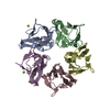
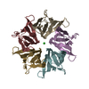
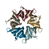
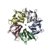
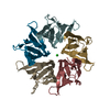
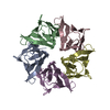
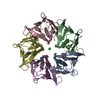
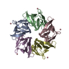
| Title | The structure and function of Xenopus NO38-core, a histone chaperone in the nucleolus | ||||||
 Components Components | Nucleophosmin NPM1 NPM1 | ||||||
 Keywords Keywords | CHAPERONE / NO38 / drosophila Nucleoplasmin-like protein (dNLP) / Nucleoplasmin (Np) / histone binding | ||||||
| Function / homology |  Function and homology informationregulation of endoribonuclease activity / regulation of endodeoxyribonuclease activity / DNA repair / nucleolus / RNA binding / nucleoplasm / identical protein binding / cytoplasm Function and homology informationregulation of endoribonuclease activity / regulation of endodeoxyribonuclease activity / DNA repair / nucleolus / RNA binding / nucleoplasm / identical protein binding / cytoplasmSimilarity search - Function | ||||||
| Biological species | Xenopus laevis (African clawed frog) | ||||||
| Method | X-RAY DIFFRACTION / MOLECULAR REPLACEMENT / Resolution: 1.9 Å | ||||||
 Authors Authors | Namboodiri, V.M. / Akey, I.V. / Schmidt-Zachmann, M.S. / Head, J.F. / Akey, C.W. | ||||||
 Citation Citation | Journal: Structure / Year: 2004 Title: The Structure and Function of Xenopus NO38-Core, a Histone Chaperone in the Nucleolus. Authors: Namboodiri, V.M. / Akey, I.V. / Schmidt-Zachmann, M.S. / Head, J.F. / Akey, C.W. | ||||||
| History |
|
- Structure visualization
| Structure viewer | Molecule: MolmilJmol/JSmol |
|---|
- Downloads & links
-Download
| PDBx/mmCIF format | 1xb9.cif.gz | 210 KB | Display | PDBx/mmCIF format |
|---|---|---|---|---|
| PDB format | pdb1xb9.ent.gz | 171.3 KB | Display | PDB format |
| PDBx/mmJSON format | 1xb9.json.gz | Tree view | PDBx/mmJSON format | |
| Others |  Other downloads Other downloads |
-Validation report
| Arichive directory | https://data.pdbj.org/pub/pdb/validation_reports/xb/1xb9ftp://data.pdbj.org/pub/pdb/validation_reports/xb/1xb9 | HTTPS FTP |
|---|
-Related structure data
| Related structure data |  1xe0C  1k5jS S: Starting model for refinement C: citing same article ( |
|---|---|
| Similar structure data |
-Links
 PDBj
PDBj

- Assembly
| Deposited unit | 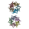
| ||||||||
|---|---|---|---|---|---|---|---|---|---|
| 1 | 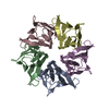
| ||||||||
| 2 | 
| ||||||||
| Unit cell |
|
-Components
| #1: Protein | NPM1 / NPM / Nucleolar phosphoprotein B23 / Numatrin / Nucleolar protein NO38 Mass: 12351.986 Da / Num. of mol.: 10 / Fragment: N-terminal core (residues 16-124) Source method: isolated from a genetically manipulated source Source: (gene. exp.) Xenopus laevis (African clawed frog) / Plasmid: pPEP-T / Species (production host): Escherichia coli / Production host:  Escherichia coli BL21(DE3) (bacteria) / Strain (production host): BL21(DE3) / References: UniProt: P07222 Escherichia coli BL21(DE3) (bacteria) / Strain (production host): BL21(DE3) / References: UniProt: P07222#2: Water | ChemComp-HOH / | Water Mass: 18.015 Da / Num. of mol.: 305 / Source method: isolated from a natural source / Formula: H2O Mass: 18.015 Da / Num. of mol.: 305 / Source method: isolated from a natural source / Formula: H2O |
|---|
-Experimental details
-Experiment
| Experiment | Method: X-RAY DIFFRACTION / Number of used crystals: 1 |
|---|
- Sample preparation
| Crystal | Density Matthews: 2.17 Å3/Da / Density % sol: 43 % |
|---|---|
| Crystal grow | Temperature: 296 K / Method: vapor diffusion, hanging drop / pH: 7.5 Details: PEG400, Tris-HCl, Calcium Chloride, pH 7.5, VAPOR DIFFUSION, HANGING DROP, temperature 296K |
-Data collection
| Diffraction | Mean temperature: 100 K |
|---|---|
| Diffraction source | Source: ROTATING ANODE / Type: RIGAKU RU300 / Wavelength: 1.54 Å |
| Detector | Type: RIGAKU RAXIS IV++ / Detector: IMAGE PLATE / Date: Oct 30, 2002 / Details: Mirrors |
| Radiation | Monochromator: Osmic Mirrors / Protocol: SINGLE WAVELENGTH / Monochromatic (M) / Laue (L): M / Scattering type: x-ray |
| Radiation wavelength | Wavelength: 1.54 Å / Relative weight: 1 |
| Reflection | Resolution: 1.9→90 Å / Num. obs: 89372 / % possible obs: 94 % / Observed criterion σ(F): 0 / Redundancy: 4.9 % / Rmerge(I) obs: 0.038 / Rsym value: 0.031 / Net I/σ(I): 14.5 |
| Reflection shell | Resolution: 1.9→1.97 Å / Redundancy: 4.2 % / Rmerge(I) obs: 0.221 / Mean I/σ(I) obs: 3.24 / Num. unique all: 369 / Rsym value: 0.19 / % possible all: 94 |
- Processing
| Software |
| ||||||||||||||||||||||||||||||||||||||||||||||||||||||||||||||||||||||||||||||||||||||||||||||||||||
|---|---|---|---|---|---|---|---|---|---|---|---|---|---|---|---|---|---|---|---|---|---|---|---|---|---|---|---|---|---|---|---|---|---|---|---|---|---|---|---|---|---|---|---|---|---|---|---|---|---|---|---|---|---|---|---|---|---|---|---|---|---|---|---|---|---|---|---|---|---|---|---|---|---|---|---|---|---|---|---|---|---|---|---|---|---|---|---|---|---|---|---|---|---|---|---|---|---|---|---|---|---|
| Refinement | Method to determine structure: MOLECULAR REPLACEMENT Starting model: PDB Entry 1K5J Resolution: 1.9→91.29 Å / Cor.coef. Fo:Fc: 0.946 / Cor.coef. Fo:Fc free: 0.918 / SU B: 4.036 / SU ML: 0.12 / Cross valid method: THROUGHOUT / σ(F): 0 / ESU R: 0.184 / ESU R Free: 0.172 / Stereochemistry target values: MAXIMUM LIKELIHOOD
| ||||||||||||||||||||||||||||||||||||||||||||||||||||||||||||||||||||||||||||||||||||||||||||||||||||
| Solvent computation | Ion probe radii: 0.8 Å / Shrinkage radii: 0.8 Å / VDW probe radii: 1.4 Å / Solvent model: BABINET MODEL WITH MASK | ||||||||||||||||||||||||||||||||||||||||||||||||||||||||||||||||||||||||||||||||||||||||||||||||||||
| Displacement parameters | Biso mean: 25.843 Å2
| ||||||||||||||||||||||||||||||||||||||||||||||||||||||||||||||||||||||||||||||||||||||||||||||||||||
| Refine analyze | Luzzati coordinate error obs: 0.29 Å / Luzzati d res low obs: 5 Å / Luzzati sigma a obs: 0.23 Å | ||||||||||||||||||||||||||||||||||||||||||||||||||||||||||||||||||||||||||||||||||||||||||||||||||||
| Refinement step | Cycle: LAST / Resolution: 1.9→91.29 Å
| ||||||||||||||||||||||||||||||||||||||||||||||||||||||||||||||||||||||||||||||||||||||||||||||||||||
| Refine LS restraints |
| ||||||||||||||||||||||||||||||||||||||||||||||||||||||||||||||||||||||||||||||||||||||||||||||||||||
| LS refinement shell | Resolution: 1.9→1.949 Å / Total num. of bins used: 20
|
