 Movie
Movie Controller


+ Open data

- Basic information
| Entry | Database: PDB / ID: 1woj | ||||||
|---|---|---|---|---|---|---|---|
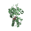
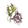
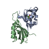
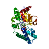
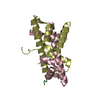
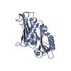
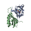
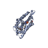
| Title | Crystal structure of human phosphodiesterase | ||||||
 Components Components | 2',3'-cyclic-nucleotide 3'-phosphodiesterase | ||||||
 Keywords Keywords | HYDROLASE | ||||||
| Function / homology |  Function and homology information Function and homology informationcyclic nucleotide catabolic process / 2',3'-cyclic-nucleotide 3'-phosphodiesterase / 2',3'-cyclic-nucleotide 3'-phosphodiesterase activity / : / myelin sheath adaxonal region / myelin sheath abaxonal region / cyclic nucleotide binding / regulation of mitochondrial membrane permeability / oligodendrocyte differentiation / pseudopodium ...cyclic nucleotide catabolic process / 2',3'-cyclic-nucleotide 3'-phosphodiesterase / 2',3'-cyclic-nucleotide 3'-phosphodiesterase activity / : / myelin sheath adaxonal region / myelin sheath abaxonal region / cyclic nucleotide binding / regulation of mitochondrial membrane permeability / oligodendrocyte differentiation / pseudopodium / microvillus / forebrain development / substantia nigra development / axonogenesis / adult locomotory behavior / response to toxic substance / microtubule cytoskeleton organization / melanosome / chemical synaptic transmission / mitochondrial inner membrane / microtubule / mitochondrial outer membrane / response to lipopolysaccharide / synapse / perinuclear region of cytoplasm / extracellular space / RNA binding / extracellular exosome / nucleoplasm / membrane / plasma membrane / cytosol / cytoplasmSimilarity search - Function | ||||||
| Biological species |  Homo sapiens (human) Homo sapiens (human) | ||||||
| Method | X-RAY DIFFRACTION / SYNCHROTRON / SIR / Resolution: 1.8 Å | ||||||
 Authors Authors | Sakamoto, Y. / Tanaka, N. / Ichimiya, T. / Kurihara, T. / Nakamura, K.T. | ||||||
 Citation Citation | Journal: J.Mol.Biol. / Year: 2005 Title: Crystal structure of the catalytic fragment of human brain 2',3'-cyclic-nucleotide 3'-phosphodiesterase Authors: Sakamoto, Y. / Tanaka, N. / Ichimiya, T. / Kurihara, T. / Nakamura, K.T. | ||||||
| History |
|
- Structure visualization
| Structure viewer | Molecule: MolmilJmol/JSmol |
|---|
- Downloads & links
-Download
| PDBx/mmCIF format | 1woj.cif.gz | 54.6 KB | Display | PDBx/mmCIF format |
|---|---|---|---|---|
| PDB format | pdb1woj.ent.gz | 39.7 KB | Display | PDB format |
| PDBx/mmJSON format | 1woj.json.gz | Tree view | PDBx/mmJSON format | |
| Others |  Other downloads Other downloads |
-Validation report
| Arichive directory | https://data.pdbj.org/pub/pdb/validation_reports/wo/1wojftp://data.pdbj.org/pub/pdb/validation_reports/wo/1woj | HTTPS FTP |
|---|
-Related structure data
| Similar structure data |
|---|


-Links
 PDBj
PDBj- Assembly
| Deposited unit | 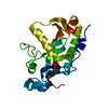
| ||||||||
|---|---|---|---|---|---|---|---|---|---|
| 1 |
| ||||||||
| Unit cell |
|
-Components
| #1: Protein | / CNP / CNPase Mass: 23946.387 Da / Num. of mol.: 1 / Fragment: residues 166-379 Source method: isolated from a genetically manipulated source Source: (gene. exp.) Homo sapiens (human) / Plasmid: pGEX-6P / Species (production host): Escherichia coli / Production host:  Escherichia coli BL21(DE3) (bacteria) / Strain (production host): BL21(DE3) Escherichia coli BL21(DE3) (bacteria) / Strain (production host): BL21(DE3)References: UniProt: P09543, 2',3'-cyclic-nucleotide 3'-phosphodiesterase |
|---|---|
| #2: Chemical | ChemComp-PO4 / Phosphate  Mass: 94.971 Da / Num. of mol.: 1 / Source method: obtained synthetically / Formula: PO4 Mass: 94.971 Da / Num. of mol.: 1 / Source method: obtained synthetically / Formula: PO4 |
| #3: Water | ChemComp-HOH / Water Mass: 18.015 Da / Num. of mol.: 96 / Source method: isolated from a natural source / Formula: H2O Mass: 18.015 Da / Num. of mol.: 96 / Source method: isolated from a natural source / Formula: H2O |
-Experimental details
-Experiment
| Experiment | Method: X-RAY DIFFRACTION / Number of used crystals: 1 |
|---|
- Sample preparation
| Crystal | Density Matthews: 1.91 Å3/Da / Density % sol: 33 % |
|---|---|
| Crystal grow | Temperature: 293 K / Method: vapor diffusion, hanging drop / pH: 5.5 Details: PEG300, sodium citrate, pH 5.5, VAPOR DIFFUSION, HANGING DROP, temperature 293K |
-Data collection
| Diffraction | Mean temperature: 100 K |
|---|---|
| Diffraction source | Source: SYNCHROTRON / Site: Photon Factory  / Beamline: AR-NW12A / Wavelength: 1 Å / Beamline: AR-NW12A / Wavelength: 1 Å |
| Detector | Type: ADSC QUANTUM 210 / Detector: CCD / Date: May 29, 2003 |
| Radiation | Protocol: SINGLE WAVELENGTH / Monochromatic (M) / Laue (L): M / Scattering type: x-ray |
| Radiation wavelength | Wavelength: 1 Å / Relative weight: 1 |
| Reflection | Resolution: 1.8→40 Å / Num. obs: 18263 / % possible obs: 98.2 % / Redundancy: 6.5 % / Rsym value: 0.057 / Net I/σ(I): 9.4 |
| Reflection shell | Resolution: 1.8→1.9 Å / Redundancy: 4.9 % / Mean I/σ(I) obs: 3.8 / Rsym value: 0.191 / % possible all: 92 |
- Processing
| Software |
| ||||||||||||||||||||||||||||||||||||||||||||||||||||||||||||||||||||||||||||||||||||||||||||||||||||
|---|---|---|---|---|---|---|---|---|---|---|---|---|---|---|---|---|---|---|---|---|---|---|---|---|---|---|---|---|---|---|---|---|---|---|---|---|---|---|---|---|---|---|---|---|---|---|---|---|---|---|---|---|---|---|---|---|---|---|---|---|---|---|---|---|---|---|---|---|---|---|---|---|---|---|---|---|---|---|---|---|---|---|---|---|---|---|---|---|---|---|---|---|---|---|---|---|---|---|---|---|---|
| Refinement | Method to determine structure: SIR / Resolution: 1.8→40 Å / Cor.coef. Fo:Fc: 0.944 / Cor.coef. Fo:Fc free: 0.929 / SU B: 2.435 / SU ML: 0.078 / Cross valid method: THROUGHOUT / ESU R: 0.146 / ESU R Free: 0.132 / Stereochemistry target values: MAXIMUM LIKELIHOOD / Details: HYDROGENS HAVE BEEN ADDED IN THE RIDING POSITIONS
| ||||||||||||||||||||||||||||||||||||||||||||||||||||||||||||||||||||||||||||||||||||||||||||||||||||
| Solvent computation | Ion probe radii: 0.8 Å / Shrinkage radii: 0.8 Å / VDW probe radii: 1.4 Å / Solvent model: BABINET MODEL WITH MASK | ||||||||||||||||||||||||||||||||||||||||||||||||||||||||||||||||||||||||||||||||||||||||||||||||||||
| Displacement parameters | Biso mean: 21.465 Å2
| ||||||||||||||||||||||||||||||||||||||||||||||||||||||||||||||||||||||||||||||||||||||||||||||||||||
| Refinement step | Cycle: LAST / Resolution: 1.8→40 Å
| ||||||||||||||||||||||||||||||||||||||||||||||||||||||||||||||||||||||||||||||||||||||||||||||||||||
| Refine LS restraints |
| ||||||||||||||||||||||||||||||||||||||||||||||||||||||||||||||||||||||||||||||||||||||||||||||||||||
| LS refinement shell | Resolution: 1.8→1.847 Å / Total num. of bins used: 20 /
|
