 Movie
Movie Controller

[English] 日本語
 Yorodumi
- PDB-1we0: Crystal structure of peroxiredoxin (AhpC) from Amphibacillus xylanus -
+ Open data

- Basic information
| Entry | Database: PDB / ID: 1we0 | ||||||
|---|---|---|---|---|---|---|---|
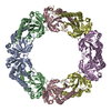
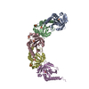
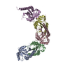
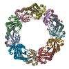
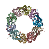
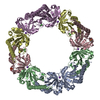
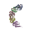
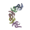
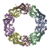
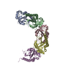
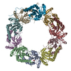
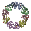
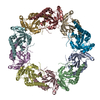
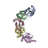
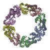
| Title | Crystal structure of peroxiredoxin (AhpC) from Amphibacillus xylanus | ||||||
 Components Components | alkyl hydroperoxide reductase C | ||||||
 Keywords Keywords |  OXIDOREDUCTASE / Peroxiredoxin / AhpC OXIDOREDUCTASE / Peroxiredoxin / AhpC | ||||||
| Function / homology |  Function and homology information Function and homology informationNADH-dependent peroxiredoxin / NADH-dependent peroxiredoxin activity / peroxiredoxin activity / peroxidase activity / response to oxidative stress / cytoplasmSimilarity search - Function | ||||||
| Biological species |  Amphibacillus xylanus (bacteria) Amphibacillus xylanus (bacteria) | ||||||
| Method | X-RAY DIFFRACTION / SYNCHROTRON / MOLECULAR REPLACEMENT / Resolution: 2.9 Å | ||||||
 Authors Authors | Kitano, K. / Kita, A. / Hakoshima, T. / Niimura, Y. / Miki, K. | ||||||
 Citation Citation | Journal: Proteins / Year: 2005 Title: Crystal structure of decameric peroxiredoxin (AhpC) from Amphibacillus xylanus Authors: Kitano, K. / Kita, A. / Hakoshima, T. / Niimura, Y. / Miki, K. #1: Journal: J.Biochem.(Tokyo) / Year: 1999 Title: Stimulation of peroxidase activity by decamerization related to ionic strength: AhpC protein from Amphibacillus xylanus Authors: Kitano, K. / Niimura, Y. / Nishiyama, Y. / Miki, K. | ||||||
| History |
|
- Structure visualization
| Structure viewer | Molecule: MolmilJmol/JSmol |
|---|
- Downloads & links
-Download
| PDBx/mmCIF format | 1we0.cif.gz | 286.8 KB | Display | PDBx/mmCIF format |
|---|---|---|---|---|
| PDB format | pdb1we0.ent.gz | 248.5 KB | Display | PDB format |
| PDBx/mmJSON format | 1we0.json.gz | Tree view | PDBx/mmJSON format | |
| Others |  Other downloads Other downloads |
-Validation report
| Arichive directory | https://data.pdbj.org/pub/pdb/validation_reports/we/1we0ftp://data.pdbj.org/pub/pdb/validation_reports/we/1we0 | HTTPS FTP |
|---|
-Related structure data
| Similar structure data |
|---|


-Links
 PDBj
PDBj
- Assembly
| Deposited unit | 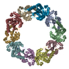
| ||||||||
|---|---|---|---|---|---|---|---|---|---|
| 1 |
| ||||||||
| Unit cell |
| ||||||||
| Details | The biological assembly is a decamer in the asymmetric unit |
-Components
| #1: Protein | Mass: 20663.092 Da / Num. of mol.: 10 Source method: isolated from a genetically manipulated source Source: (gene. exp.) Amphibacillus xylanus (bacteria) / Plasmid: pAHNO2.5 / Production host: Escherichia coli (E. coli) / Strain (production host): JM109References: UniProt: O87200, UniProt: K0J4Q8*PLUS, Oxidoreductases; Acting on a peroxide as acceptor; Peroxidases#2: Chemical | Ammonium  Mass: 18.038 Da / Num. of mol.: 2 / Source method: obtained synthetically / Formula: H4N Mass: 18.038 Da / Num. of mol.: 2 / Source method: obtained synthetically / Formula: H4N |
|---|
-Experimental details
-Experiment
| Experiment | Method: X-RAY DIFFRACTION / Number of used crystals: 2 |
|---|
- Sample preparation
| Crystal | Density Matthews: 3 Å3/Da / Density % sol: 59.49 % |
|---|---|
| Crystal grow | Temperature: 293 K / Method: vapor diffusion, hanging drop / pH: 5.5 Details: PEG 6000, pH 5.5, VAPOR DIFFUSION, HANGING DROP, temperature 293K |
-Data collection
| Diffraction |
| |||||||||||||||
|---|---|---|---|---|---|---|---|---|---|---|---|---|---|---|---|---|
| Diffraction source |
| |||||||||||||||
| Detector |
| |||||||||||||||
| Radiation | Protocol: SINGLE WAVELENGTH / Monochromatic (M) / Laue (L): M / Scattering type: x-ray | |||||||||||||||
| Radiation wavelength | Wavelength: 1 Å / Relative weight: 1 | |||||||||||||||
| Reflection | Resolution: 2.9→50 Å / Num. obs: 46092 / % possible obs: 83.6 % / Observed criterion σ(I): 12.5 | |||||||||||||||
| Reflection shell | Resolution: 2.9→3 Å / % possible all: 55.1 |

- Processing
| Software |
| ||||||||||||
|---|---|---|---|---|---|---|---|---|---|---|---|---|---|
| Refinement | Method to determine structure: MOLECULAR REPLACEMENT / Resolution: 2.9→50 Å / σ(F): 1
| ||||||||||||
| Refinement step | Cycle: LAST / Resolution: 2.9→50 Å
|
