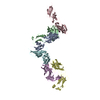
 Movie
Movie Controller


+ Open data

- Basic information
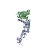
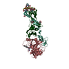
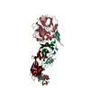
| Entry | Database: PDB / ID: 1w0y | ||||||
|---|---|---|---|---|---|---|---|
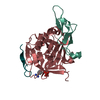
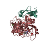
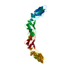
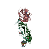
| Title | tf7a_3771 complex | ||||||
 Components Components |
| ||||||
 Keywords Keywords |  HYDROLASE / SERINE PROTEASE / BLOOD COAGULATION / GLYCOPROTEIN / PLASMA / VITAMIN K / CALCIUM-BINDING / GAMMA-CARBOXYGLUTAMIC ACID / CO-FACTOR / COAGULATION / ENZYME COMPLEX HYDROLASE / SERINE PROTEASE / BLOOD COAGULATION / GLYCOPROTEIN / PLASMA / VITAMIN K / CALCIUM-BINDING / GAMMA-CARBOXYGLUTAMIC ACID / CO-FACTOR / COAGULATION / ENZYME COMPLEX | ||||||
| Function / homology |  Function and homology information Function and homology informationactivation of plasma proteins involved in acute inflammatory response / activation of blood coagulation via clotting cascade / coagulation factor VIIa / response to Thyroid stimulating hormone / response to 2,3,7,8-tetrachlorodibenzodioxine / response to astaxanthin / response to thyrotropin-releasing hormone / response to genistein / serine-type peptidase complex / positive regulation of platelet-derived growth factor receptor signaling pathway ...activation of plasma proteins involved in acute inflammatory response / activation of blood coagulation via clotting cascade / coagulation factor VIIa / response to Thyroid stimulating hormone / response to 2,3,7,8-tetrachlorodibenzodioxine / response to astaxanthin / response to thyrotropin-releasing hormone / response to genistein / serine-type peptidase complex / positive regulation of platelet-derived growth factor receptor signaling pathway / response to vitamin K / response to carbon dioxide / response to thyroxine / positive regulation of leukocyte chemotaxis / NGF-stimulated transcription / response to cholesterol / response to growth hormone / cytokine receptor activity / positive regulation of positive chemotaxis / Extrinsic Pathway of Fibrin Clot Formation / positive regulation of TOR signaling / positive regulation of blood coagulation / animal organ regeneration / Gamma-carboxylation of protein precursors / Transport of gamma-carboxylated protein precursors from the endoplasmic reticulum to the Golgi apparatus / Removal of aminoterminal propeptides from gamma-carboxylated proteins / positive regulation of endothelial cell proliferation / serine-type peptidase activity / BMAL1:CLOCK,NPAS2 activates circadian gene expression / positive regulation of interleukin-8 production / phospholipid binding / protein processing / Golgi lumen / cytokine-mediated signaling pathway / circadian rhythm / response to estrogen / activation of cysteine-type endopeptidase activity involved in apoptotic process / positive regulation of angiogenesis / blood coagulation / response to estradiol / collagen-containing extracellular matrix / protease binding / vesicle / response to hypoxia / positive regulation of cell migration / endoplasmic reticulum lumen / external side of plasma membrane / signaling receptor binding / serine-type endopeptidase activity / calcium ion binding / positive regulation of gene expression / cell surface / extracellular space / extracellular region / membrane / plasma membraneSimilarity search - Function | ||||||
| Biological species |  HOMO SAPIENS (human) HOMO SAPIENS (human) | ||||||
| Method | X-RAY DIFFRACTION / SYNCHROTRON / MOLECULAR REPLACEMENT / Resolution: 2.5 Å | ||||||
 Authors Authors | Banner, D.W. / D'Arcy, A. / Groebke-Zbinden, K. / Ackermann, J. / Kirchhofer, D. / Ji, Y.-H. / Tschopp, T.B. / Wallbaum, S. / Weber, L. | ||||||
 Citation Citation | Journal: Bioorg.Med.Chem.Lett. / Year: 2005 Title: Design of Selective Phenylglycine Amide Tissue Factor/Factor Viia Inhibitors Authors: Groebke-Zbinden, K. / Banner, D.W. / Ackermann, J. / D'Arcy, A. / Kirchhofer, D. / Ji, Y.-H. / Tschopp, T.B. / Wallbaum, S. / Weber, L. #1: Journal: Nature / Year: 1996Title: The Crystal Structure of the Complex of Blood Coagulation Factor Viia with Soluble Tissue Factor Authors: Banner, D.W. / D'Arcy, A. / Chene, C. / Winkler, F.K. / Guha, A. / Konigsberg, W.H. / Nemerson, Y. / Kirchhofer, D. | ||||||
| History |
| ||||||
| Remark 700 | SHEET DETERMINATION METHOD: DSSP THE SHEETS PRESENTED AS "HA HB" IN EACH CHAIN ON SHEET RECORDS ... SHEET DETERMINATION METHOD: DSSP THE SHEETS PRESENTED AS "HA HB" IN EACH CHAIN ON SHEET RECORDS BELOW IS ACTUALLY AN 8-STRANDED BARREL THIS IS REPRESENTED BY A 9-STRANDED SHEET IN WHICH THE FIRST AND LAST STRANDS ARE IDENTICAL. |
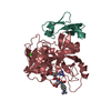
- Structure visualization
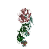
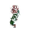
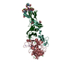
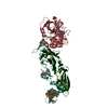
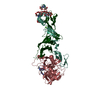
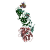
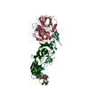
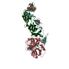
| Structure viewer | Molecule: MolmilJmol/JSmol |
|---|
- Downloads & links
-Download
| PDBx/mmCIF format | 1w0y.cif.gz | 147.4 KB | Display | PDBx/mmCIF format |
|---|---|---|---|---|
| PDB format | pdb1w0y.ent.gz | 111.6 KB | Display | PDB format |
| PDBx/mmJSON format | 1w0y.json.gz | Tree view | PDBx/mmJSON format | |
| Others |  Other downloads Other downloads |
-Validation report
| Arichive directory | https://data.pdbj.org/pub/pdb/validation_reports/w0/1w0yftp://data.pdbj.org/pub/pdb/validation_reports/w0/1w0y | HTTPS FTP |
|---|
-Related structure data
| Related structure data |  1w2kC  1danS S: Starting model for refinement C: citing same article ( |
|---|---|
| Similar structure data |


-Links
 PDBj
PDBj


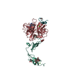
- Assembly
| Deposited unit | 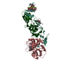
| ||||||||
|---|---|---|---|---|---|---|---|---|---|
| 1 |
| ||||||||
| Unit cell |
| ||||||||
| Details | THIS DESIGNATION IS BASED ON AN AUTOMATIC QUATERNARYSTRUCTURE DETERMINATION (PQS.EBI.AC.UK). |
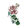
-Components
-BLOOD COAGULATION FACTOR ... , 2 types, 2 molecules HL
| #1: Protein | Mass: 28103.256 Da / Num. of mol.: 1 / Fragment: FACTOR VII HEAVY CHAIN, RESIDUES 213-466 Source method: isolated from a genetically manipulated source Source: (gene. exp.) HOMO SAPIENS (human)Cell line (production host): BABY HAMSTER KIDNEY CELLS (BHK) Production host:   CRICETULUS GRISEUS (Chinese hamster) / References: UniProt: P08709, coagulation factor VIIa CRICETULUS GRISEUS (Chinese hamster) / References: UniProt: P08709, coagulation factor VIIa |
|---|---|
| #2: Protein | Mass: 16315.761 Da / Num. of mol.: 1 / Fragment: FACTOR VII HEAVY CHAIN, RESIDUES 61-202 Source method: isolated from a genetically manipulated source Source: (gene. exp.) HOMO SAPIENS (human)Cell line (production host): BABY HAMSTER KIDNEY CELLS (BHK) Production host: CRICETULUS GRISEUS (Chinese hamster) / References: UniProt: P08709, coagulation factor VIIa |
-Protein , 1 types, 1 molecules T
| #3: Protein | / TF / COAGULATION FACTOR III / THROMBOPLASTIN / CD142 ANTIGEN / SOLUBLE TISSUE FACTOR Mass: 23763.393 Da / Num. of mol.: 1 / Fragment: EXTRACELLULAR DOMAIN, RESIDUES 38-242 Source method: isolated from a genetically manipulated source Source: (gene. exp.) HOMO SAPIENS (human) / Production host:  ESCHERICHIA COLI (E. coli) / References: UniProt: P13726 ESCHERICHIA COLI (E. coli) / References: UniProt: P13726 |
|---|
-Sugars , 2 types, 2 molecules 

| #7: Sugar | ChemComp-FUC / Fucose Type: L-saccharide, alpha linking / Mass: 164.156 Da / Num. of mol.: 1 Type: L-saccharide, alpha linking / Mass: 164.156 Da / Num. of mol.: 1Source method: isolated from a genetically manipulated source Formula: C6H12O5 |
|---|---|
| #8: Sugar | ChemComp-BGC / Glucose Type: D-saccharide, beta linking / Mass: 180.156 Da / Num. of mol.: 1 Type: D-saccharide, beta linking / Mass: 180.156 Da / Num. of mol.: 1Source method: isolated from a genetically manipulated source Formula: C6H12O6 |
-Non-polymers , 4 types, 384 molecules 


| #4: Chemical | ChemComp-771 /  Mass: 454.542 Da / Num. of mol.: 1 / Source method: obtained synthetically / Formula: C23H26N4O4S Mass: 454.542 Da / Num. of mol.: 1 / Source method: obtained synthetically / Formula: C23H26N4O4S | ||||
|---|---|---|---|---|---|
| #5: Chemical | ChemComp-CA /  Mass: 40.078 Da / Num. of mol.: 9 / Source method: obtained synthetically / Formula: Ca Mass: 40.078 Da / Num. of mol.: 9 / Source method: obtained synthetically / Formula: Ca#6: Chemical | ChemComp-CAC / | Cacodylic acid Mass: 136.989 Da / Num. of mol.: 1 / Source method: obtained synthetically / Formula: C2H6AsO2 Mass: 136.989 Da / Num. of mol.: 1 / Source method: obtained synthetically / Formula: C2H6AsO2#9: Water | ChemComp-HOH / | WaterMass: 18.015 Da / Num. of mol.: 373 / Source method: isolated from a natural source / Formula: H2O |
-Details
| Compound details | THE PEPTIDE GROUP LINKING RESIDUES 192 AND 193 OF THE HEAVY CHAIN OF FACTOR VIIA IS FLIPPED 180 ...THE PEPTIDE GROUP LINKING RESIDUES 192 AND 193 OF THE HEAVY CHAIN OF FACTOR VIIA IS FLIPPED 180 DEGREES FROM THE STANDARD ACTIVE CONFORMATI |
|---|
-Experimental details
-Experiment
| Experiment | Method: X-RAY DIFFRACTION / Number of used crystals: 1 |
|---|
- Sample preparation
| Crystal | Density Matthews: 2.8 Å3/Da / Density % sol: 49.5 % / Description: 1DAN WAS ROOM TEMPERATURE - THIS IS FROZEN |
|---|---|
| Crystal grow | pH: 5.5 / Details: pH 5.50 |
-Data collection
| Diffraction | Mean temperature: 100 K |
|---|---|
| Diffraction source | Source: SYNCHROTRON / Site: ESRF  / Beamline: BM1A / Wavelength: 0.873 / Beamline: BM1A / Wavelength: 0.873 |
| Detector | Type: MARRESEARCH / Detector: IMAGE PLATE / Date: Jan 30, 1997 |
| Radiation | Protocol: SINGLE WAVELENGTH / Monochromatic (M) / Laue (L): M / Scattering type: x-ray |
| Radiation wavelength | Wavelength: 0.873 Å / Relative weight: 1 |
| Reflection | Resolution: 2.46→25 Å / Num. obs: 85076 / % possible obs: 87.8 % / Observed criterion σ(I): 0 / Redundancy: 3.27 % / Biso Wilson estimate: 35.4 Å2 / Rmerge(I) obs: 0.13 |
| Reflection shell | Resolution: 2.46→2.58 Å / Redundancy: 2.6 % / Rmerge(I) obs: 0.39 / % possible all: 50.3 |
- Processing
| Software |
| ||||||||||||||||||||||||||||||||||||||||||||||||||||||||||||||||||||||||||||||||
|---|---|---|---|---|---|---|---|---|---|---|---|---|---|---|---|---|---|---|---|---|---|---|---|---|---|---|---|---|---|---|---|---|---|---|---|---|---|---|---|---|---|---|---|---|---|---|---|---|---|---|---|---|---|---|---|---|---|---|---|---|---|---|---|---|---|---|---|---|---|---|---|---|---|---|---|---|---|---|---|---|---|
| Refinement | Method to determine structure: MOLECULAR REPLACEMENT Starting model: PDB ENTRY 1DAN Resolution: 2.5→19.99 Å / Rfactor Rfree error: 0.007 / Isotropic thermal model: RESTRAINED / Cross valid method: THROUGHOUT / σ(F): 0
| ||||||||||||||||||||||||||||||||||||||||||||||||||||||||||||||||||||||||||||||||
| Solvent computation | Solvent model: FLAT MODEL / Bsol: 45.3164 Å2 / ksol: 0.340884 e/Å3 | ||||||||||||||||||||||||||||||||||||||||||||||||||||||||||||||||||||||||||||||||
| Displacement parameters | Biso mean: 22.7 Å2
| ||||||||||||||||||||||||||||||||||||||||||||||||||||||||||||||||||||||||||||||||
| Refine analyze |
| ||||||||||||||||||||||||||||||||||||||||||||||||||||||||||||||||||||||||||||||||
| Refinement step | Cycle: LAST / Resolution: 2.5→19.99 Å
| ||||||||||||||||||||||||||||||||||||||||||||||||||||||||||||||||||||||||||||||||
| Refine LS restraints |
| ||||||||||||||||||||||||||||||||||||||||||||||||||||||||||||||||||||||||||||||||
| LS refinement shell | Resolution: 2.5→2.59 Å / Rfactor Rfree error: 0.028 / Total num. of bins used: 10
| ||||||||||||||||||||||||||||||||||||||||||||||||||||||||||||||||||||||||||||||||
| Xplor file |
|
