 Movie
Movie Controller


+ Open data

- Basic information
| Entry | Database: PDB / ID: 1w0j | ||||||
|---|---|---|---|---|---|---|---|
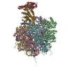
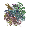
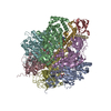
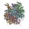
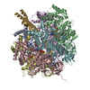
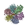
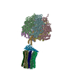
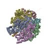
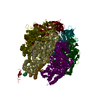
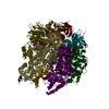
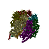
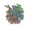
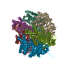
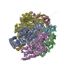
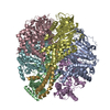
| Title | Beryllium fluoride inhibited bovine F1-ATPase | ||||||
 Components Components | (ATP SYNTHASE ... ) x 3 ) x 3 | ||||||
 Keywords Keywords | HYDROLASE / ATP PHOSPHORYLASE / ATP PHOSPHORYLASE (H+ TRANSPORTING) / ATP SYNTHASE / F1FO ATP SYNTHASE / F1-ATPASE / ATP SYNTHESIS / ATP-BINDING | ||||||
| Function / homology |  Function and homology information Function and homology informationFormation of ATP by chemiosmotic coupling / Cristae formation / mitochondrial proton-transporting ATP synthase, catalytic core / mitochondrial proton-transporting ATP synthase complex / mitochondrial proton-transporting ATP synthase complex, catalytic sector F(1) / proton motive force-driven mitochondrial ATP synthesis / proton motive force-driven ATP synthesis / proton-transporting ATP synthase complex, catalytic core F(1) / H+-transporting two-sector ATPase / proton-transporting ATPase activity, rotational mechanism ...Formation of ATP by chemiosmotic coupling / Cristae formation / mitochondrial proton-transporting ATP synthase, catalytic core / mitochondrial proton-transporting ATP synthase complex / mitochondrial proton-transporting ATP synthase complex, catalytic sector F(1) / proton motive force-driven mitochondrial ATP synthesis / proton motive force-driven ATP synthesis / proton-transporting ATP synthase complex, catalytic core F(1) / H+-transporting two-sector ATPase / proton-transporting ATPase activity, rotational mechanism / proton-transporting ATP synthase activity, rotational mechanism / ADP binding / ATP hydrolysis activity / ATP binding / plasma membraneSimilarity search - Function | ||||||
| Biological species |  BOS TAURUS (cattle) BOS TAURUS (cattle) | ||||||
| Method | X-RAY DIFFRACTION / SYNCHROTRON / MOLECULAR REPLACEMENT / Resolution: 2.2 Å | ||||||
 Authors Authors | Kagawa, R. / Montgomery, M.G. / Braig, K. / Walker, J.E. / Leslie, A.G.W. | ||||||
 Citation Citation | Journal: Embo J. / Year: 2004 Title: The Structure of Bovine F1-ATPase Inhibited by Adp and Beryllium Fluoride Authors: Kagawa, R. / Montgomery, M.G. / Braig, K. / Leslie, A.G.W. / Walker, J.E. #1: Journal: Cell(Cambridge,Mass.) / Year: 2001Title: Structure of Bovine Mitochondrial F1-ATPase with Nucleotide Bound to All Three Catalytic Sites; Implications for the Mechanism of Rotary Catalysis Authors: Menz, R.I. / Walker, J.E. / Leslie, A.G.W. #2: Journal: Nat.Struct.Biol. / Year: 2000Title: The Structure of the Central Stalk in Bovine F1-ATPase at 2.4A Resolution Authors: Gibbons, C. / Montgomery, M.G. / Leslie, A.G.W. / Walker, J.E. #3: Journal: Science / Year: 1999Title: Molecular Architecture of the Rotary Motor in ATP Synthase Authors: Stock, D. / Leslie, A.G.W. / Walker, J.E. #4: Journal: Angew.Chem.Int.Ed.Engl. / Year: 1998Title: ATP Synthesis by Rotary Catalysis (Nobel Lecture) Authors: Walker, J.E. #5: Journal: Nature / Year: 1994Title: Structure at 2.8 A Resolution of F1-ATPase from Bovine Heart Mitochondria Authors: Abrahams, J.P. / Leslie, A.G.W. / Lutter, R. / Walker, J.E. #6: Journal: J.Mol.Biol. / Year: 1993 Title: Crystallization of F1-ATPase from Bovine Heart Mitochondria. Authors: Lutter, R. / Abrahams, J.P. / Van Raaij, M.J. / Todd, R.J. / Lundqvist, T. / Buchanan, S.K. / Leslie, A.G. / Walker, J.E. | ||||||
| History |
| ||||||
| Remark 700 | SHEET DETERMINATION METHOD: DSSP THE SHEETS PRESENTED AS "AA" IN EACH CHAIN ON SHEET RECORDS BELOW ... SHEET DETERMINATION METHOD: DSSP THE SHEETS PRESENTED AS "AA" IN EACH CHAIN ON SHEET RECORDS BELOW IS ACTUALLY AN 13-STRANDED BARREL THIS IS REPRESENTED BY A 14-STRANDED SHEET IN WHICH THE FIRST AND LAST STRANDS ARE IDENTICAL. THE SHEETS PRESENTED AS "BA" IN EACH CHAIN ON SHEET RECORDS BELOW IS ACTUALLY AN 11-STRANDED BARREL THIS IS REPRESENTED BY A 12-STRANDED SHEET IN WHICH THE FIRST AND LAST STRANDS ARE IDENTICAL. THE SHEETS PRESENTED AS "CA" IN EACH CHAIN ON SHEET RECORDS BELOW IS ACTUALLY AN 11-STRANDED BARREL THIS IS REPRESENTED BY A 12-STRANDED SHEET IN WHICH THE FIRST AND LAST STRANDS ARE IDENTICAL. THE SHEET STRUCTURE OF THIS MOLECULE IS BIFURCATED. IN ORDER TO REPRESENT THIS FEATURE IN THE SHEET RECORDS BELOW, TWO SHEETS ARE DEFINED. |
- Structure visualization
| Structure viewer | Molecule: MolmilJmol/JSmol |
|---|
- Downloads & links
-Download
| PDBx/mmCIF format | 1w0j.cif.gz | 609.1 KB | Display | PDBx/mmCIF format |
|---|---|---|---|---|
| PDB format | pdb1w0j.ent.gz | 495.9 KB | Display | PDB format |
| PDBx/mmJSON format | 1w0j.json.gz | Tree view | PDBx/mmJSON format | |
| Others |  Other downloads Other downloads |
-Validation report
| Arichive directory | https://data.pdbj.org/pub/pdb/validation_reports/w0/1w0jftp://data.pdbj.org/pub/pdb/validation_reports/w0/1w0j | HTTPS FTP |
|---|
-Related structure data
| Related structure data |  1w0kC  1e1qS S: Starting model for refinement C: citing same article ( |
|---|---|
| Similar structure data |


-Links
 PDBj
PDBj


- Assembly
| Deposited unit | 
| ||||||||
|---|---|---|---|---|---|---|---|---|---|
| 1 |
| ||||||||
| Unit cell |
|
-Components
-ATP SYNTHASE ... , 3 types, 7 molecules ABCDEFG
| #1: Protein | Mass: 55301.207 Da / Num. of mol.: 3 / Source method: isolated from a natural source / Source: (natural) BOS TAURUS (cattle) / Organ: HEART / Tissue: MUSCLESkeletal muscleReferences: UniProt: P19483, H+-transporting two-sector ATPase#2: Protein | Mass: 51757.836 Da / Num. of mol.: 3 / Source method: isolated from a natural source / Source: (natural) BOS TAURUS (cattle) / Organ: HEART / Tissue: MUSCLESkeletal muscleReferences: UniProt: P00829, H+-transporting two-sector ATPase#3: Protein | | Mass: 30185.674 Da / Num. of mol.: 1 / Source method: isolated from a natural source / Source: (natural) BOS TAURUS (cattle) / Organ: HEART / Tissue: MUSCLESkeletal muscleReferences: UniProt: P05631, H+-transporting two-sector ATPase |
|---|
-Non-polymers , 6 types, 1054 molecules 


| #4: Chemical | ChemComp-ADP / Adenosine diphosphate Mass: 427.201 Da / Num. of mol.: 5 / Source method: obtained synthetically / Formula: C10H15N5O10P2 / Comment: ADP, energy-carrying molecule*YM Mass: 427.201 Da / Num. of mol.: 5 / Source method: obtained synthetically / Formula: C10H15N5O10P2 / Comment: ADP, energy-carrying molecule*YM#5: Chemical | ChemComp-MG /  Mass: 24.305 Da / Num. of mol.: 5 / Source method: obtained synthetically / Formula: Mg Mass: 24.305 Da / Num. of mol.: 5 / Source method: obtained synthetically / Formula: Mg#6: Chemical | ChemComp-GOL / Glycerol Mass: 92.094 Da / Num. of mol.: 4 / Source method: obtained synthetically / Formula: C3H8O3 Mass: 92.094 Da / Num. of mol.: 4 / Source method: obtained synthetically / Formula: C3H8O3#7: Chemical |  Mass: 66.007 Da / Num. of mol.: 2 / Source method: obtained synthetically / Formula: BeF3 Mass: 66.007 Da / Num. of mol.: 2 / Source method: obtained synthetically / Formula: BeF3#8: Chemical | ChemComp-PO4 / | Phosphate Mass: 94.971 Da / Num. of mol.: 1 / Source method: obtained synthetically / Formula: PO4 Mass: 94.971 Da / Num. of mol.: 1 / Source method: obtained synthetically / Formula: PO4#9: Water | ChemComp-HOH / | WaterMass: 18.015 Da / Num. of mol.: 1037 / Source method: isolated from a natural source / Formula: H2O |
|---|
-Details
| Compound details | THE F1-ATPASE MOLECULE HAS THREE COPIES OF THE NON-CATALYTIC ALPHA SUBUNIT AND THREE COPIES OF THE ...THE F1-ATPASE MOLECULE HAS THREE COPIES OF THE NON-CATALYTIC ALPHA SUBUNIT AND THREE COPIES OF THE CATALYTIC BETA SUBUNIT. IN THE 1994 REFERENCE, THE BETA SUBUNITS WERE LABELED ACCORDING TO THE BOUND NUCLEOTIDE |
|---|---|
| Sequence details | REFERENCE: 1) FOR THE ALPHA SUBUNIT: J.E.WALKER,S.J.POWELL, O.VINAS AND M.J.RUNSWICK, BIOCHEMISTRY ...REFERENCE: 1) FOR THE ALPHA SUBUNIT: J.E.WALKER,S.J.POWELL, O.VINAS AND M.J.RUNSWICK, BIOCHEMIST |
-Experimental details
-Experiment
| Experiment | Method: X-RAY DIFFRACTION / Number of used crystals: 1 |
|---|
- Sample preparation
| Crystal | Density Matthews: 2.99 Å3/Da / Density % sol: 54 % |
|---|---|
| Crystal grow | pH: 8.2 Details: CRYSTALS WERE GROWN IN THE PRESENCE OF AZIDE, A KNOWN INHIBITOR, BUT THIS HAS NOT BEEN LOCATED IN THE STRUCTURE., pH 8.20 |
-Data collection
| Diffraction | Mean temperature: 100 K |
|---|---|
| Diffraction source | Source: SYNCHROTRON / Site: ESRF  / Beamline: ID14-4 / Wavelength: 0.94 / Beamline: ID14-4 / Wavelength: 0.94 |
| Detector | Type: ADSC CCD / Detector: CCD / Date: Mar 15, 2002 |
| Radiation | Protocol: SINGLE WAVELENGTH / Monochromatic (M) / Laue (L): M / Scattering type: x-ray |
| Radiation wavelength | Wavelength: 0.94 Å / Relative weight: 1 |
| Reflection | Resolution: 2.2→45.2 Å / Num. obs: 185113 / % possible obs: 87.1 % / Observed criterion σ(I): 0 / Redundancy: 2 % / Rmerge(I) obs: 0.08 / Net I/σ(I): 8.1 |
| Reflection shell | Resolution: 2.2→2.32 Å / Redundancy: 1.4 % / Rmerge(I) obs: 0.354 / Mean I/σ(I) obs: 1.7 / % possible all: 51.1 |
- Processing
| Software |
| ||||||||||||||||||||||||||||||||||||||||||||||||||||||||||||||||||||||||||||||||||||||||||||||||||||||||||||||||||||||||||||||||||||||||||||||||||
|---|---|---|---|---|---|---|---|---|---|---|---|---|---|---|---|---|---|---|---|---|---|---|---|---|---|---|---|---|---|---|---|---|---|---|---|---|---|---|---|---|---|---|---|---|---|---|---|---|---|---|---|---|---|---|---|---|---|---|---|---|---|---|---|---|---|---|---|---|---|---|---|---|---|---|---|---|---|---|---|---|---|---|---|---|---|---|---|---|---|---|---|---|---|---|---|---|---|---|---|---|---|---|---|---|---|---|---|---|---|---|---|---|---|---|---|---|---|---|---|---|---|---|---|---|---|---|---|---|---|---|---|---|---|---|---|---|---|---|---|---|---|---|---|---|---|---|---|
| Refinement | Method to determine structure: MOLECULAR REPLACEMENT Starting model: PDB CODE 1E1Q, NATIVE BOVINE MITOCHONDRIAL F1- ATPASE Resolution: 2.2→20 Å / Cor.coef. Fo:Fc: 0.956 / Cor.coef. Fo:Fc free: 0.93 / Cross valid method: THROUGHOUT / Stereochemistry target values: MAXIMUM LIKELIHOOD Details: THE PHOSPHATE GROUP ADJACENT TO THE P-LOOP OF THE BETA(E) SUBUNIT (CHAIN E) IN THE COORDINATES HAS A HIGH B FACTOR AND DOES NOT HAVE THE EXPECTED HYDROGEN BONDS TO THE PROTEIN. IT IS ...Details: THE PHOSPHATE GROUP ADJACENT TO THE P-LOOP OF THE BETA(E) SUBUNIT (CHAIN E) IN THE COORDINATES HAS A HIGH B FACTOR AND DOES NOT HAVE THE EXPECTED HYDROGEN BONDS TO THE PROTEIN. IT IS POSSIBLE THAT THIS IS NOT, IN FACT, A PHOSPHATE, AND IT PROBABLY DOES NOT REPRESENT A PHYSIOLOGICALLY RELEVANT PHOSPHATE BINDING SITE.
| ||||||||||||||||||||||||||||||||||||||||||||||||||||||||||||||||||||||||||||||||||||||||||||||||||||||||||||||||||||||||||||||||||||||||||||||||||
| Solvent computation | Ion probe radii: 0.8 Å / Shrinkage radii: 0.8 Å / VDW probe radii: 1.2 Å / Solvent model: BABINET MODEL WITH MASK | ||||||||||||||||||||||||||||||||||||||||||||||||||||||||||||||||||||||||||||||||||||||||||||||||||||||||||||||||||||||||||||||||||||||||||||||||||
| Displacement parameters | Biso mean: 48.59 Å2
| ||||||||||||||||||||||||||||||||||||||||||||||||||||||||||||||||||||||||||||||||||||||||||||||||||||||||||||||||||||||||||||||||||||||||||||||||||
| Refinement step | Cycle: LAST / Resolution: 2.2→20 Å
| ||||||||||||||||||||||||||||||||||||||||||||||||||||||||||||||||||||||||||||||||||||||||||||||||||||||||||||||||||||||||||||||||||||||||||||||||||
| Refine LS restraints |
|