 Movie
Movie Controller


+ Open data

- Basic information
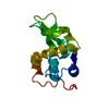
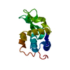
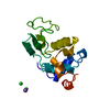
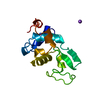
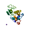
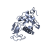
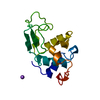
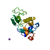
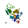
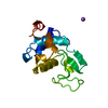
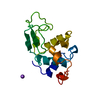
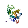
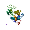
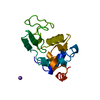
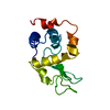
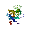
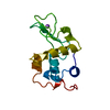
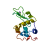
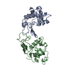
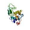
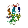
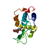
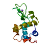
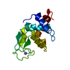
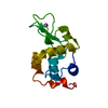
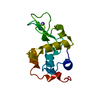
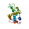
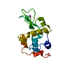
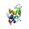
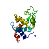
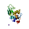
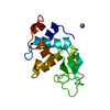
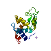
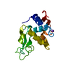
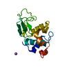
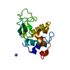
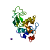
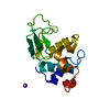
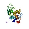
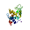
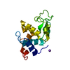
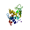
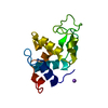
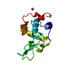
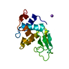
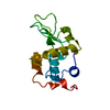
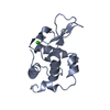
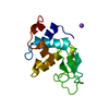
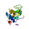
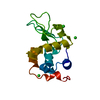
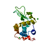
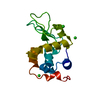
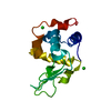
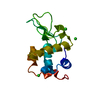
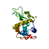
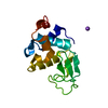
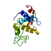
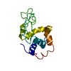
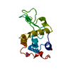
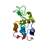
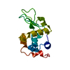
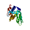
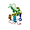
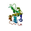
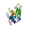
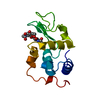
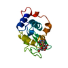
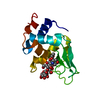
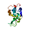
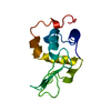
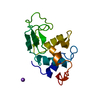
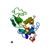
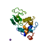
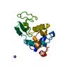
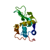
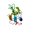
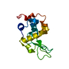
| Entry | Database: PDB / ID: 1w08 | ||||||
|---|---|---|---|---|---|---|---|
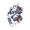
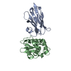
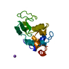
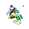
| Title | STRUCTURE OF T70N HUMAN LYSOZYME | ||||||
 Components Components | LYSOZYME | ||||||
 Keywords Keywords | HYDROLASE / O-GLYCOSYL / HUMAN LYSOZYME / ENZYME / AMYLOID | ||||||
| Function / homology |  Function and homology informationcytolysis / antimicrobial humoral response / retina homeostasis / Antimicrobial peptides / metabolic process / specific granule lumen / azurophil granule lumen / tertiary granule lumen / lysozyme / lysozyme activity ...cytolysis / antimicrobial humoral response / retina homeostasis / Antimicrobial peptides / metabolic process / specific granule lumen / azurophil granule lumen / tertiary granule lumen / lysozyme / lysozyme activity / killing of cells of another organism / defense response to Gram-negative bacterium / defense response to Gram-positive bacterium / defense response to bacterium / inflammatory response / Amyloid fiber formation / Neutrophil degranulation / extracellular space / extracellular exosome / extracellular region / identical protein binding Function and homology informationcytolysis / antimicrobial humoral response / retina homeostasis / Antimicrobial peptides / metabolic process / specific granule lumen / azurophil granule lumen / tertiary granule lumen / lysozyme / lysozyme activity ...cytolysis / antimicrobial humoral response / retina homeostasis / Antimicrobial peptides / metabolic process / specific granule lumen / azurophil granule lumen / tertiary granule lumen / lysozyme / lysozyme activity / killing of cells of another organism / defense response to Gram-negative bacterium / defense response to Gram-positive bacterium / defense response to bacterium / inflammatory response / Amyloid fiber formation / Neutrophil degranulation / extracellular space / extracellular exosome / extracellular region / identical protein bindingSimilarity search - Function | ||||||
| Biological species |  HOMO SAPIENS (human) HOMO SAPIENS (human) | ||||||
| Method | X-RAY DIFFRACTION / MOLECULAR REPLACEMENT / Resolution: 2.5 Å | ||||||
 Authors Authors | Johnson, R. / Christodoulou, J. / Luisi, B. / Dumoulin, M. / Caddy, G. / Alcocer, M. / Murtagh, G. / Archer, D.B. / Dobson, C.M. | ||||||
 Citation Citation | Journal: J.Mol.Biol. / Year: 2005 Title: Rationalising Lysozyme Amyloidosis: Insights from the Structure and Solution Dynamics of T70N Lysozyme. Authors: Johnson, R. / Christodoulou, J. / Dumoulin, M. / Caddy, G. / Alcocer, M. / Murtagh, G. / Kumita, J.R. / Larsson, G. / Robinson, C.V. / Archer, D.B. / Luisi, B. / Dobson, C.M. | ||||||
| History |
|
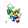
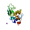
- Structure visualization
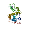
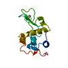
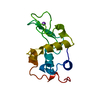
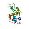
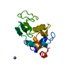
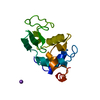
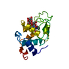
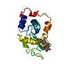
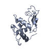
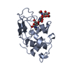
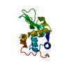
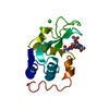
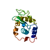
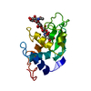
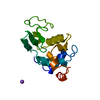
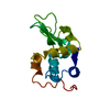
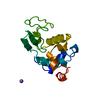
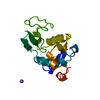
| Structure viewer | Molecule: MolmilJmol/JSmol |
|---|
- Downloads & links
-Download
| PDBx/mmCIF format | 1w08.cif.gz | 43.1 KB | Display | PDBx/mmCIF format |
|---|---|---|---|---|
| PDB format | pdb1w08.ent.gz | 30.1 KB | Display | PDB format |
| PDBx/mmJSON format | 1w08.json.gz | Tree view | PDBx/mmJSON format | |
| Others |  Other downloads Other downloads |
-Validation report
| Arichive directory | https://data.pdbj.org/pub/pdb/validation_reports/w0/1w08ftp://data.pdbj.org/pub/pdb/validation_reports/w0/1w08 | HTTPS FTP |
|---|
-Related structure data
| Related structure data | 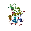 1jsfS S: Starting model for refinement |
|---|---|
| Similar structure data |


-Links
 PDBj
PDBj


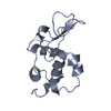
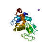
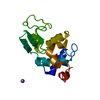
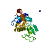
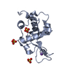
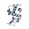
- Assembly
| Deposited unit | 
| ||||||||
|---|---|---|---|---|---|---|---|---|---|
| 1 |
| ||||||||
| Unit cell |
|
-Components
| #1: Protein | / 1 / 4-BETA-N-ACETYLMURAMIDASE C Mass: 14733.691 Da / Num. of mol.: 1 / Fragment: RESIDUES 19-148 Source method: isolated from a genetically manipulated source Source: (gene. exp.) HOMO SAPIENS (human) / Production host:  PICHIA PASTORIS (fungus) / Strain (production host): GS 115 / References: UniProt: P00695, UniProt: P61626*PLUS, lysozyme PICHIA PASTORIS (fungus) / Strain (production host): GS 115 / References: UniProt: P00695, UniProt: P61626*PLUS, lysozyme |
|---|---|
| #2: Chemical | ChemComp-CL / Chloride  Mass: 35.453 Da / Num. of mol.: 1 / Source method: obtained synthetically / Formula: Cl Mass: 35.453 Da / Num. of mol.: 1 / Source method: obtained synthetically / Formula: Cl |
| #3: Water | ChemComp-HOH / Water Mass: 18.015 Da / Num. of mol.: 88 / Source method: isolated from a natural source / Formula: H2O Mass: 18.015 Da / Num. of mol.: 88 / Source method: isolated from a natural source / Formula: H2O |
| Sequence details | VARIANT DESCRIBED IN UNIPROT WITH FTID=VAR_012050 IN P00695 |
-Experimental details
-Experiment
| Experiment | Method: X-RAY DIFFRACTION / Number of used crystals: 1 |
|---|
- Sample preparation
| Crystal | Density Matthews: 1.86 Å3/Da / Density % sol: 33 % |
|---|---|
| Crystal grow | Temperature: 293 K / Method: vapor diffusion, hanging drop / pH: 4.5 Details: HANGING DROP METHOD AT 293K. DROPLET RESERVOIR SOLUTION MIXED 1:1 WITH 10 MG/ML PROTEIN, 10 MM HEPES BUFFER PH 7.5, 0.4 M LICL. RESERVOIR 2.5 M NACL, 20 MM NAOAC PH 4.5. |
-Data collection
| Diffraction | Mean temperature: 100 K |
|---|---|
| Diffraction source | Source: ROTATING ANODE / Type: RIGAKU RU200 / Wavelength: 1.5418 |
| Detector | Type: MSC / Detector: IMAGE PLATE / Date: Nov 15, 2004 / Details: OSMIC MIRROR |
| Radiation | Monochromator: OSMIC MIRROR / Protocol: SINGLE WAVELENGTH / Monochromatic (M) / Laue (L): M / Scattering type: x-ray |
| Radiation wavelength | Wavelength: 1.5418 Å / Relative weight: 1 |
| Reflection | Resolution: 2.5→20 Å / Num. obs: 3903 / % possible obs: 95.1 % / Redundancy: 6.4 % / Rmerge(I) obs: 0.075 / Net I/σ(I): 14.2 |
| Reflection shell | Resolution: 2.5→2.59 Å / Redundancy: 2.9 % / Rmerge(I) obs: 0.146 / Mean I/σ(I) obs: 6.5 / % possible all: 87.6 |
- Processing
| Software |
| ||||||||||||||||||||
|---|---|---|---|---|---|---|---|---|---|---|---|---|---|---|---|---|---|---|---|---|---|
| Refinement | Method to determine structure: MOLECULAR REPLACEMENT Starting model: PDB ENTRY 1JSF Resolution: 2.5→41.89 Å / SU B: 14.331 / SU ML: 0.332 / Cross valid method: THROUGHOUT / ESU R Free: 0.399 / Details: HYDROGENS ADDED IN RIDING POSITIONS.
| ||||||||||||||||||||
| Displacement parameters | Biso mean: 23.657 Å2
| ||||||||||||||||||||
| Refinement step | Cycle: LAST / Resolution: 2.5→41.89 Å
|
