 Movie
Movie Controller


+ Open data

- Basic information
| Entry | Database: PDB / ID: 1vwc | ||||||
|---|---|---|---|---|---|---|---|
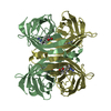
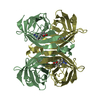
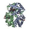
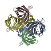
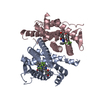
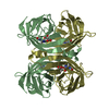
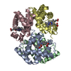
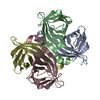
| Title | STREPTAVIDIN-CYCLO-AC-[CHPQFC]-NH2, PH 2.0 | ||||||
 Components Components |
| ||||||
 Keywords Keywords | COMPLEX (BIOTIN-BINDING PROTEIN/PEPTIDE) / COMPLEX (BIOTIN-BINDING PROTEIN-PEPTIDE) / CYCLIC PEPTIDE DISCOVERED BY PHAGE DISPLAY / COMPLEX (BIOTIN-BINDING PROTEIN-PEPTIDE) complex | ||||||
| Function / homology |  Function and homology information Function and homology information | ||||||
| Biological species |   Streptomyces avidinii (bacteria) Streptomyces avidinii (bacteria) | ||||||
| Method | X-RAY DIFFRACTION / Resolution: 1.86 Å | ||||||
 Authors Authors | Katz, B.A. / Cass, R.T. | ||||||
 Citation Citation | Journal: J.Biol.Chem. / Year: 1997 Title: In crystals of complexes of streptavidin with peptide ligands containing the HPQ sequence the pKa of the peptide histidine is less than 3.0. Authors: Katz, B.A. / Cass, R.T. #1: Journal: J.Am.Chem.Soc. / Year: 1996Title: Structure-Based Design Tools: Structural and Thermodynamic Comparison with Biotin of a Small Molecule that Binds Streptavidin with Micromolar Affinity Authors: Katz, B.A. / Liu, B. / Cass, R.T. #2: Journal: J.Am.Chem.Soc. / Year: 1996Title: Preparation of a Protein-Dimerizing Ligand by Topochemistry and Structure-Based Design Authors: Katz, B.A. #3: Journal: J.Biol.Chem. / Year: 1995Title: Topochemical Catalysis Achieved by Structure-Based Ligand Design Authors: Katz, B.A. / Cass, R.T. / Liu, B. / Arze, R. / Collins, N. #4: Journal: Chem.Biol. / Year: 1995Title: Topochemistry for Preparing Ligands that Dimerize Receptors Authors: Katz, B.A. / Stroud, R.M. / Collins, N. / Liu, B. / Arze, R. #5: Journal: Biochemistry / Year: 1995Title: Binding to Protein Targets of Peptidic Leads Discovered by Phage Display: Crystal Structures of Streptavidin-Bound Linear and Cyclic Peptide Ligands Containing the Hpq Sequence Authors: Katz, B.A. #6: Journal: J.Am.Chem.Soc. / Year: 1995Title: Structure-Based Design of High Affinity Streptavidin Binding Cyclic Peptide Ligands Containing Thioether Cross-Links Authors: Katz, B.A. / Johnson, C.R. / Cass, R.T. | ||||||
| History |
|
- Structure visualization
| Structure viewer | Molecule: MolmilJmol/JSmol |
|---|
- Downloads & links
-Download
| PDBx/mmCIF format | 1vwc.cif.gz | 44.2 KB | Display | PDBx/mmCIF format |
|---|---|---|---|---|
| PDB format | pdb1vwc.ent.gz | 35 KB | Display | PDB format |
| PDBx/mmJSON format | 1vwc.json.gz | Tree view | PDBx/mmJSON format | |
| Others |  Other downloads Other downloads |
-Validation report
| Arichive directory | https://data.pdbj.org/pub/pdb/validation_reports/vw/1vwcftp://data.pdbj.org/pub/pdb/validation_reports/vw/1vwc | HTTPS FTP |
|---|
-Related structure data
| Related structure data | 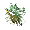 1vwaC 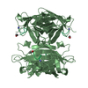 1vwbC 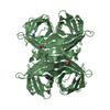 1vwdC 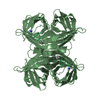 1vweC 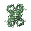 1vwfC 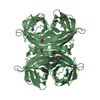 1vwgC 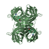 1vwhC 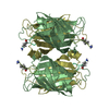 1vwiC 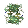 1vwjC 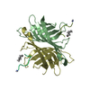 1vwkC 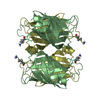 1vwlC  1vwmC 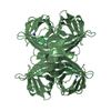 1vwnC 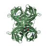 1vwoC  1vwpC 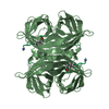 1vwqC 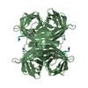 1vwrC C: citing same article ( |
|---|---|
| Similar structure data |


-Links
 PDBj
PDBj- Assembly
| Deposited unit | 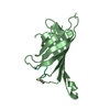
| ||||||||
|---|---|---|---|---|---|---|---|---|---|
| 1 | 
| ||||||||
| Unit cell |
|
-Components
| #1: Protein | Mass: 12965.025 Da / Num. of mol.: 1 Source method: isolated from a genetically manipulated source Source: (gene. exp.) Streptomyces avidinii (bacteria) / References: UniProt: P22629 |
|---|---|
| #2: Protein/peptide | Mass: 758.911 Da / Num. of mol.: 1 Source method: isolated from a genetically manipulated source |
| #3: Water | ChemComp-HOH / Water Mass: 18.015 Da / Num. of mol.: 65 / Source method: isolated from a natural source / Formula: H2O Mass: 18.015 Da / Num. of mol.: 65 / Source method: isolated from a natural source / Formula: H2O |
-Experimental details
-Experiment
| Experiment | Method: X-RAY DIFFRACTION / Number of used crystals: 1 |
|---|
- Sample preparation
| Crystal | Density Matthews: 2.81 Å3/Da / Density % sol: 36 % | |||||||||||||||||||||||||||||||||||||||||||||||||
|---|---|---|---|---|---|---|---|---|---|---|---|---|---|---|---|---|---|---|---|---|---|---|---|---|---|---|---|---|---|---|---|---|---|---|---|---|---|---|---|---|---|---|---|---|---|---|---|---|---|---|
| Crystal grow | pH: 2 Details: SYNTHETIC MOTHER LIQUOR = 75 % SATURATED AMMONIUM SULFATE, 25 % 1.0 M SODIUM FORMATE ADJUSTED TO PH 2.0. | |||||||||||||||||||||||||||||||||||||||||||||||||
| Crystal grow | *PLUS Temperature: 20 ℃ / pH: 4.5 / Method: vapor diffusion, hanging drop / Details: Pahler, A., (1987) J. Biol. Chem., 262, 13933. | |||||||||||||||||||||||||||||||||||||||||||||||||
| Components of the solutions | *PLUS
|
-Data collection
| Diffraction | Mean temperature: 295 K |
|---|---|
| Diffraction source | Wavelength: 1.5418 |
| Detector | Type: SIEMENS / Detector: AREA DETECTOR |
| Radiation | Monochromatic (M) / Laue (L): M / Scattering type: x-ray |
| Radiation wavelength | Wavelength: 1.5418 Å / Relative weight: 1 |
| Reflection | Num. obs: 12424 / Redundancy: 3.9 % / Rmerge(I) obs: 0.053 |
| Reflection | *PLUS Highest resolution: 1.79 Å / Num. measured all: 49532 |
- Processing
| Software |
| ||||||||||||||||||||||||||||||||||||||||||||||||||||||||||||
|---|---|---|---|---|---|---|---|---|---|---|---|---|---|---|---|---|---|---|---|---|---|---|---|---|---|---|---|---|---|---|---|---|---|---|---|---|---|---|---|---|---|---|---|---|---|---|---|---|---|---|---|---|---|---|---|---|---|---|---|---|---|
| Refinement | Resolution: 1.86→7.5 Å / σ(F): 2 Details: THE FOLLOWING ATOMS HAD WEAK DENSITY AND OCCUPANCIES WERE REFINED: 13, 14, 15, SIDE CHAIN OF GLN 24, TERMINUS OF ASP 36, SIDE CHAIN OF 47, 48, 50, 51, TERMINUS OF ARG 53, 64, 65, 66, 67, 99, ...Details: THE FOLLOWING ATOMS HAD WEAK DENSITY AND OCCUPANCIES WERE REFINED: 13, 14, 15, SIDE CHAIN OF GLN 24, TERMINUS OF ASP 36, SIDE CHAIN OF 47, 48, 50, 51, TERMINUS OF ARG 53, 64, 65, 66, 67, 99, 100, SIDE CHAIN OF 101, TERMINUS OF ARG 103, TERMINUS OF GLU 116, TERMINUS OF LYS 121, (ACE P 0 AND CYS P 1), (CYS P 6 AND NH2 P 7). DISCRETELY DISORDERED SIDE CHAINS WHOSE OCCUPANCIES AND STRUCTURES WERE SIMULTANEOUSLY REFINED WERE 43, 84 (3 CONFORMATIONS), 107, 132. DISORDERED WATERS ARE HOH 207 WHICH IS CLOSE TO A SYMMETRY-RELATED EQUIVALENT OF HOH 354; HOH 244 WHICH IS CLOSE TO A SYMMETRY-RELATED EQUIVALENT OF HOH 298. HOH 305, HOH 313, AND HOH 467 ARE CLOSE TO SYMMETRY-RELATED EQUIVALENTS OF THEMSELVES, RESPECTIVELY. NO ENERGY INTERACTIONS INVOLVING HOH 305, HOH 313, OR HOH 467 WERE TURNED ON DURING REFINEMENT. THE FOLLOWING ATOMS HAD WEAK DENSITY AND OCCUPANCIES WERE REFINED: 13, 14, 15, SIDE CHAIN OF GLN 24, TERMINUS OF ASP 36, SIDE CHAIN OF 47, 48, 50, 51, TERMINUS OF ARG 53, 64, 65, 66, 67, 99, 100, SIDE CHAIN OF 101, TERMINUS OF ARG 103, TERMINUS OF GLU 116, TERMINUS OF LYS 121, (ACE P 0 AND CYS P 1), (CYS P 6 AND NH2 P 7).
| ||||||||||||||||||||||||||||||||||||||||||||||||||||||||||||
| Refinement step | Cycle: LAST / Resolution: 1.86→7.5 Å
| ||||||||||||||||||||||||||||||||||||||||||||||||||||||||||||
| Refine LS restraints |
| ||||||||||||||||||||||||||||||||||||||||||||||||||||||||||||
| LS refinement shell | Resolution: 1.86→1.94 Å / % reflection obs: 28.9 % | ||||||||||||||||||||||||||||||||||||||||||||||||||||||||||||
| Xplor file |
| ||||||||||||||||||||||||||||||||||||||||||||||||||||||||||||
| Software | *PLUS Name: X-PLOR / Classification: refinement | ||||||||||||||||||||||||||||||||||||||||||||||||||||||||||||
| Refine LS restraints | *PLUS
|
