 Movie
Movie Controller


+ Open data

- Basic information
| Entry | Database: PDB / ID: 1vdl | ||||||
|---|---|---|---|---|---|---|---|
| Title | Solution Structure of RSGI RUH-013, a UBA domain in Mouse cDNA | ||||||
 Components Components | Ubiquitin carboxyl-terminal hydrolase 25 | ||||||
 Keywords Keywords |  STRUCTURAL GENOMICS / UNKNOWN FUNCTION / UBA domain / Mouse cDNA / RIKEN Structural Genomics/Proteomics Initiative / RSGI STRUCTURAL GENOMICS / UNKNOWN FUNCTION / UBA domain / Mouse cDNA / RIKEN Structural Genomics/Proteomics Initiative / RSGI | ||||||
| Function / homology |  Function and homology information Function and homology informationnegative regulation of ERAD pathway / SUMO binding / protein K48-linked deubiquitination / Ub-specific processing proteases / protein K63-linked deubiquitination / protein deubiquitination / proteasome complex / ATPase binding / ubiquitin-dependent protein catabolic process / ubiquitinyl hydrolase 1 ...negative regulation of ERAD pathway / SUMO binding / protein K48-linked deubiquitination / Ub-specific processing proteases / protein K63-linked deubiquitination / protein deubiquitination / proteasome complex / ATPase binding / ubiquitin-dependent protein catabolic process / ubiquitinyl hydrolase 1 / cysteine-type deubiquitinase activity / cysteine-type endopeptidase activity / ubiquitin protein ligase binding / endoplasmic reticulum / nucleus / cytosolSimilarity search - Function | ||||||
| Biological species |  Mus musculus (house mouse) Mus musculus (house mouse) | ||||||
| Method | SOLUTION NMR / torsion angle dynamics | ||||||
 Authors Authors | Doi-Katayama, Y. / Hirota, H. / Saito, K. / Koshiba, S. / Kigawa, T. / Yokoyama, S. / RIKEN Structural Genomics/Proteomics Initiative (RSGI) | ||||||
 Citation Citation | Journal: To be Published Title: Solution Structure of RSGI RUH-013, a UBA domain in Mouse cDNA Authors: Doi-Katayama, Y. / Hirota, H. / Saito, K. / Koshiba, S. / Kigawa, T. / Yokoyama, S. | ||||||
| History |
|
- Structure visualization
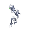
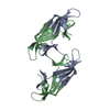
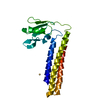
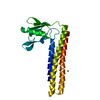
| Structure viewer | Molecule: MolmilJmol/JSmol |
|---|
- Downloads & links
-Download
| PDBx/mmCIF format | 1vdl.cif.gz | 453.8 KB | Display | PDBx/mmCIF format |
|---|---|---|---|---|
| PDB format | pdb1vdl.ent.gz | 380.4 KB | Display | PDB format |
| PDBx/mmJSON format | 1vdl.json.gz | Tree view | PDBx/mmJSON format | |
| Others |  Other downloads Other downloads |
-Validation report
| Arichive directory | https://data.pdbj.org/pub/pdb/validation_reports/vd/1vdlftp://data.pdbj.org/pub/pdb/validation_reports/vd/1vdl | HTTPS FTP |
|---|
-Related structure data
| Similar structure data | |
|---|---|
| Other databases |


-Links
 PDBj
PDBj


- Assembly
| Deposited unit | 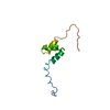
| |||||||||
|---|---|---|---|---|---|---|---|---|---|---|
| 1 |
| |||||||||
| NMR ensembles |
|
-Components
| #1: Protein | Mass: 8417.175 Da / Num. of mol.: 1 / Fragment: UBA domain Source method: isolated from a genetically manipulated source Source: (gene. exp.) Mus musculus (house mouse) / Description: Cell-free protein synthesis (E.coli) / Gene: RIKEN cDNA 2610101O11 / Plasmid: P030324-44 / References: UniProt: P57080, EC: 3.1.2.15 |
|---|
-Experimental details
-Experiment
| Experiment | Method: SOLUTION NMR | ||||||||||||
|---|---|---|---|---|---|---|---|---|---|---|---|---|---|
| NMR experiment |
|
- Sample preparation
| Details | Contents: 0.3mM UBA domain U-15N, 13C; 20mM phosphate buffer (pH 6.0); 100mM NaCl; 1mM DTT/0.02% NaN3; 90% H2O, 10% D2O Solvent system: 90% H2O/10% D2O |
|---|---|
| Sample conditions | Ionic strength: 120mM NaCl / pH: 6.0 / Pressure: ambient / Temperature: 298 K |
-NMR measurement
| Radiation | Protocol: SINGLE WAVELENGTH / Monochromatic (M) / Laue (L): M |
|---|---|
| Radiation wavelength | Relative weight: 1 |
| NMR spectrometer | Type: Bruker AVANCE / Manufacturer: Bruker / Model: AVANCE / Field strength: 800 MHz |
- Processing
| NMR software |
| ||||||||||||||||||||||||||||
|---|---|---|---|---|---|---|---|---|---|---|---|---|---|---|---|---|---|---|---|---|---|---|---|---|---|---|---|---|---|
| Refinement | Method: torsion angle dynamics / Software ordinal: 1 | ||||||||||||||||||||||||||||
| NMR ensemble | Conformer selection criteria: structures with the least restraint violations, structures with the lowest energy, target function Conformers calculated total number: 100 / Conformers submitted total number: 20 |
