 Movie
Movie Controller


+ Open data

- Basic information
| Entry | Database: PDB / ID: 1vb7 | ||||||
|---|---|---|---|---|---|---|---|
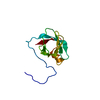
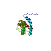
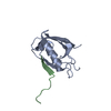
| Title | Solution structure of the PDZ domain of PDZ and LIM domain 2 | ||||||
 Components Components | PDZ and LIM domain 2 | ||||||
 Keywords Keywords |  STRUCTURAL GENOMICS / UNKNOWN FUNCTION / PDZ domain PDZ-LIM protein / RIKEN Structural Genomics/Proteomics Initiative / RSGI STRUCTURAL GENOMICS / UNKNOWN FUNCTION / PDZ domain PDZ-LIM protein / RIKEN Structural Genomics/Proteomics Initiative / RSGI | ||||||
| Function / homology |  Function and homology information Function and homology informationmuscle structure development / filamin binding / muscle alpha-actinin binding / cortical actin cytoskeleton / myosin heavy chain binding / alpha-actinin binding / filamentous actin / stress fiber / adherens junction / Z disc ...muscle structure development / filamin binding / muscle alpha-actinin binding / cortical actin cytoskeleton / myosin heavy chain binding / alpha-actinin binding / filamentous actin / stress fiber / adherens junction / Z disc / actin binding / heart development / actin cytoskeleton organization / metal ion bindingSimilarity search - Function | ||||||
| Biological species |  Mus musculus (house mouse) Mus musculus (house mouse) | ||||||
| Method | SOLUTION NMR | ||||||
 Authors Authors | Hatta, R. / Hayashi, F. / Yoshida, M. / Yokoyama, S. / RIKEN Structural Genomics/Proteomics Initiative (RSGI) | ||||||
 Citation Citation | Journal: To be Published Title: Solution structure of the PDZ domain of PDZ and LIM domain 2 Authors: Hatta, R. / Hayashi, F. / Yoshida, M. / Yokoyama, S. | ||||||
| History |
|
- Structure visualization
| Structure viewer | Molecule: MolmilJmol/JSmol |
|---|
- Downloads & links
-Download
| PDBx/mmCIF format | 1vb7.cif.gz | 527.1 KB | Display | PDBx/mmCIF format |
|---|---|---|---|---|
| PDB format | pdb1vb7.ent.gz | 442.2 KB | Display | PDB format |
| PDBx/mmJSON format | 1vb7.json.gz | Tree view | PDBx/mmJSON format | |
| Others |  Other downloads Other downloads |
-Validation report
| Arichive directory | https://data.pdbj.org/pub/pdb/validation_reports/vb/1vb7ftp://data.pdbj.org/pub/pdb/validation_reports/vb/1vb7 | HTTPS FTP |
|---|
-Related structure data
| Similar structure data | |
|---|---|
| Other databases |


-Links
 PDBj
PDBj- Assembly
| Deposited unit | 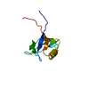
| |||||||||
|---|---|---|---|---|---|---|---|---|---|---|
| 1 |
| |||||||||
| NMR ensembles |
|
-Components
| #1: Protein | Mass: 9707.788 Da / Num. of mol.: 1 / Fragment: PDZ domain Source method: isolated from a genetically manipulated source Source: (gene. exp.) Mus musculus (house mouse) / Description: Cell-free protein synthesis / Gene: cDNA 2310041E13 / Plasmid: P030203-89 / References: UniProt: Q8R1G6 |
|---|
-Experimental details
-Experiment
| Experiment | Method: SOLUTION NMR | ||||||||||||
|---|---|---|---|---|---|---|---|---|---|---|---|---|---|
| NMR experiment |
|
- Sample preparation
| Details | Contents: 1.28mM 13C, 15N-labeled protein; 20mM phosphate buffer; 100mM NaCl; 1mM d10-DTT; 0.02% NaN3; 10% D2O Solvent system: 90% H2O/10% D2O |
|---|---|
| Sample conditions | Ionic strength: 120 / pH: 6.0 / Pressure: ambient / Temperature: 298 K |
-NMR measurement
| Radiation | Protocol: SINGLE WAVELENGTH / Monochromatic (M) / Laue (L): M |
|---|---|
| Radiation wavelength | Relative weight: 1 |
| NMR spectrometer | Type: Varian INOVA / Manufacturer: Varian / Model: INOVA / Field strength: 800 MHz |
- Processing
| NMR software |
| ||||||||||||||||||||||||||||
|---|---|---|---|---|---|---|---|---|---|---|---|---|---|---|---|---|---|---|---|---|---|---|---|---|---|---|---|---|---|
| NMR representative | Selection criteria: lowest energy | ||||||||||||||||||||||||||||
| NMR ensemble | Conformer selection criteria: structures with the least restraint violations, target function Conformers calculated total number: 100 / Conformers submitted total number: 20 |
