 Movie
Movie Controller

[English] 日本語
 Yorodumi
- PDB-1uzp: Integrin binding cbEGF22-TB4-cbEGF33 fragment of human fibrillin-... -
+ Open data

- Basic information
| Entry | Database: PDB / ID: 1uzp | ||||||
|---|---|---|---|---|---|---|---|
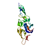
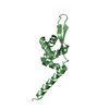
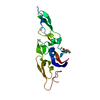
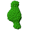
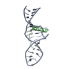
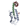
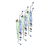
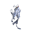
| Title | Integrin binding cbEGF22-TB4-cbEGF33 fragment of human fibrillin-1, Sm bound form cbEGF23 domain only. | ||||||
 Components Components | FIBRILLIN-1 | ||||||
 Keywords Keywords | MATRIX PROTEIN / EXTRA-CELLULAR MATRIX / FIBRILLIN-1 / CBEGF DOMAIN / TB DOMAIN | ||||||
| Function / homology |  Function and homology information Function and homology informationpost-embryonic eye morphogenesis / extracellular matrix constituent conferring elasticity / sequestering of BMP in extracellular matrix / sequestering of TGFbeta in extracellular matrix / microfibril / embryonic eye morphogenesis / negative regulation of osteoclast development / metanephros development / Elastic fibre formation / camera-type eye development ...post-embryonic eye morphogenesis / extracellular matrix constituent conferring elasticity / sequestering of BMP in extracellular matrix / sequestering of TGFbeta in extracellular matrix / microfibril / embryonic eye morphogenesis / negative regulation of osteoclast development / metanephros development / Elastic fibre formation / camera-type eye development / Molecules associated with elastic fibres / cellular response to insulin-like growth factor stimulus / cell adhesion mediated by integrin / extracellular matrix structural constituent / negative regulation of osteoclast differentiation / TGF-beta receptor signaling activates SMADs / basement membrane / anatomical structure morphogenesis / Integrin cell surface interactions / cellular response to transforming growth factor beta stimulus / extracellular matrix / Degradation of the extracellular matrix / skeletal system development / Post-translational protein phosphorylation / hormone activity / Regulation of Insulin-like Growth Factor (IGF) transport and uptake by Insulin-like Growth Factor Binding Proteins (IGFBPs) / integrin binding / heparin binding / heart development / collagen-containing extracellular matrix / endoplasmic reticulum lumen / calcium ion binding / protein-containing complex binding / extracellular space / extracellular region / identical protein bindingSimilarity search - Function | ||||||
| Biological species |  HOMO SAPIENS (human) HOMO SAPIENS (human) | ||||||
| Method | X-RAY DIFFRACTION / OTHER / Resolution: 1.78 Å | ||||||
 Authors Authors | Lee, S.S.J. / Knott, V. / Harlos, K. / Handford, P.A. / Stuart, D.I. | ||||||
 Citation Citation | Journal: Structure / Year: 2004 Title: Structure of the Integrin Binding Fragment from Fibrillin-1 Gives New Insights Into Microfibril Organization Authors: Lee, S.S.J. / Knott, V. / Jovanovi, J. / Harlos, K. / Grimes, J. / Choulier, L. / Mardon, H. / Stuart, D.I. / Handford, P.A. | ||||||
| History |
|
- Structure visualization
| Structure viewer | Molecule: MolmilJmol/JSmol |
|---|
- Downloads & links
-Download
| PDBx/mmCIF format | 1uzp.cif.gz | 44.5 KB | Display | PDBx/mmCIF format |
|---|---|---|---|---|
| PDB format | pdb1uzp.ent.gz | 33.4 KB | Display | PDB format |
| PDBx/mmJSON format | 1uzp.json.gz | Tree view | PDBx/mmJSON format | |
| Others |  Other downloads Other downloads |
-Validation report
| Arichive directory | https://data.pdbj.org/pub/pdb/validation_reports/uz/1uzpftp://data.pdbj.org/pub/pdb/validation_reports/uz/1uzp | HTTPS FTP |
|---|
-Related structure data


-Links
 PDBj
PDBj


- Assembly
| Deposited unit | 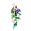
| ||||||||
|---|---|---|---|---|---|---|---|---|---|
| 1 |
| ||||||||
| Unit cell |
|
-Components
| #1: Protein | Mass: 17332.436 Da / Num. of mol.: 1 / Fragment: BEGF22-TB4-CBEGF23, RESIDUES 1486-1647 Source method: isolated from a genetically manipulated source Source: (gene. exp.) HOMO SAPIENS (human) / Tissue: EXTRA-ELLULAR MATRIX / Production host:  ESCHERICHIA COLI (E. coli) / Strain (production host): NM554 / References: UniProt: P35555 ESCHERICHIA COLI (E. coli) / Strain (production host): NM554 / References: UniProt: P35555 | ||
|---|---|---|---|
| #2: Chemical | Samarium  Mass: 150.360 Da / Num. of mol.: 3 / Source method: obtained synthetically / Formula: Sm Mass: 150.360 Da / Num. of mol.: 3 / Source method: obtained synthetically / Formula: Sm#3: Water | ChemComp-HOH / | Water Mass: 18.015 Da / Num. of mol.: 188 / Source method: isolated from a natural source / Formula: H2O Mass: 18.015 Da / Num. of mol.: 188 / Source method: isolated from a natural source / Formula: H2O |
-Experimental details
-Experiment
| Experiment | Method: X-RAY DIFFRACTION / Number of used crystals: 1 |
|---|
- Sample preparation
| Crystal | Density Matthews: 2.04 Å3/Da / Density % sol: 36 % |
|---|---|
| Crystal grow | Method: vapor diffusion, sitting drop / pH: 8.5 Details: SITTING DROP VAPOUR DIFFUSION, 3 MICROLITRE OF 25MG/ML PROTEIN, 10MM TRIS PH7.5 + 0.5 MICROLITRES OF 40% V/V POLYPROPYLENE GLYCOL P400 + 2.5 MICROLITRES RESERVOIR SOLUTION (0.2M LISULFATE, 0. ...Details: SITTING DROP VAPOUR DIFFUSION, 3 MICROLITRE OF 25MG/ML PROTEIN, 10MM TRIS PH7.5 + 0.5 MICROLITRES OF 40% V/V POLYPROPYLENE GLYCOL P400 + 2.5 MICROLITRES RESERVOIR SOLUTION (0.2M LISULFATE, 0.1M TRIS PH 8.5, 30% PEG 4000) SOAKED OVERNIGHT IN 10MM SAMARIUM ACETATE |
-Data collection
| Diffraction | Mean temperature: 100 K |
|---|---|
| Diffraction source | Source: ROTATING ANODE / Type: RIGAKU RU345 / Wavelength: 1.542 |
| Detector | Type: MARRESEARCH 300MM / Detector: IMAGE PLATE / Date: Sep 15, 2001 / Details: OSMIC BLUE CONFOCAL |
| Radiation | Protocol: SINGLE WAVELENGTH / Monochromatic (M) / Laue (L): M / Scattering type: x-ray |
| Radiation wavelength | Wavelength: 1.542 Å / Relative weight: 1 |
| Reflection | Resolution: 1.78→30 Å / Num. obs: 13997 / % possible obs: 99.5 % / Redundancy: 25.2 % / Rmerge(I) obs: 0.127 / Net I/σ(I): 24.4 |
| Reflection shell | Resolution: 1.78→1.84 Å / Rmerge(I) obs: 0.479 / Mean I/σ(I) obs: 5.3 / % possible all: 98.6 |
- Processing
| Software |
| ||||||||||||||||||||||||||||||||||||||||||||||||||||||||||||||||||||||||||||||||
|---|---|---|---|---|---|---|---|---|---|---|---|---|---|---|---|---|---|---|---|---|---|---|---|---|---|---|---|---|---|---|---|---|---|---|---|---|---|---|---|---|---|---|---|---|---|---|---|---|---|---|---|---|---|---|---|---|---|---|---|---|---|---|---|---|---|---|---|---|---|---|---|---|---|---|---|---|---|---|---|---|---|
| Refinement | Method to determine structure: OTHER / Resolution: 1.78→20 Å / Data cutoff high absF: 10000 / Isotropic thermal model: RESTRAINED / Cross valid method: THROUGHOUT / σ(F): 0
| ||||||||||||||||||||||||||||||||||||||||||||||||||||||||||||||||||||||||||||||||
| Solvent computation | Solvent model: FLAT MODEL / Bsol: 49 Å2 / ksol: 0.365 e/Å3 | ||||||||||||||||||||||||||||||||||||||||||||||||||||||||||||||||||||||||||||||||
| Displacement parameters |
| ||||||||||||||||||||||||||||||||||||||||||||||||||||||||||||||||||||||||||||||||
| Refinement step | Cycle: LAST / Resolution: 1.78→20 Å
| ||||||||||||||||||||||||||||||||||||||||||||||||||||||||||||||||||||||||||||||||
| Refine LS restraints |
| ||||||||||||||||||||||||||||||||||||||||||||||||||||||||||||||||||||||||||||||||
| LS refinement shell | Resolution: 1.78→1.8 Å / Total num. of bins used: 27 /
|
