 Movie
Movie Controller


+ Open data

- Basic information
| Entry | Database: PDB / ID: 1uyw | ||||||
|---|---|---|---|---|---|---|---|
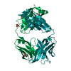
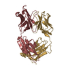
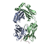
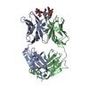
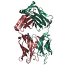
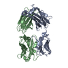
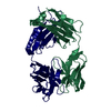
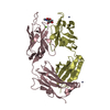
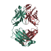
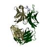
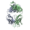
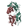
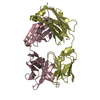
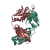
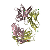
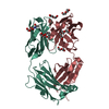
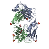
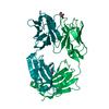
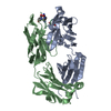
| Title | Crystal Structure of the antiflavivirus Fab4g2 | ||||||
 Components Components |
| ||||||
 Keywords Keywords |  IMMUNE SYSTEM IMMUNE SYSTEM | ||||||
| Function / homology | Immunoglobulins / Immunoglobulin-like / Sandwich / Mainly Beta Function and homology information Function and homology information | ||||||
| Biological species |  MUS MUSCULUS (house mouse) MUS MUSCULUS (house mouse) | ||||||
| Method | X-RAY DIFFRACTION / SYNCHROTRON / MOLECULAR REPLACEMENT / Resolution: 2 Å | ||||||
 Authors Authors | Martinez-Fleites, C. / Ortiz-Lombardia, M. / Taylor, E.J. / Gil-Valdes, J. / Chinea, G. / Davies, G. | ||||||
 Citation Citation | Journal: To be Published Title: Crystal Structure of the Antiflavivirus Fab4G2 Authors: Martinez-Fleites, C. / Ortiz-Lombardia, M. / Taylor, E.J. / Gil-Valdes, J. / Chinea, G. / Davies, G. | ||||||
| History |
| ||||||
| Remark 700 | SHEET THE SHEET STRUCTURE OF THIS MOLECULE IS BIFURCATED. IN ORDER TO REPRESENT THIS FEATURE IN ... SHEET THE SHEET STRUCTURE OF THIS MOLECULE IS BIFURCATED. IN ORDER TO REPRESENT THIS FEATURE IN THE SHEET RECORDS BELOW, TWO SHEETS ARE DEFINED. |
- Structure visualization
| Structure viewer | Molecule: MolmilJmol/JSmol |
|---|
- Downloads & links
-Download
| PDBx/mmCIF format | 1uyw.cif.gz | 180.3 KB | Display | PDBx/mmCIF format |
|---|---|---|---|---|
| PDB format | pdb1uyw.ent.gz | 144.8 KB | Display | PDB format |
| PDBx/mmJSON format | 1uyw.json.gz | Tree view | PDBx/mmJSON format | |
| Others |  Other downloads Other downloads |
-Validation report
| Arichive directory | https://data.pdbj.org/pub/pdb/validation_reports/uy/1uywftp://data.pdbj.org/pub/pdb/validation_reports/uy/1uyw | HTTPS FTP |
|---|
-Related structure data
| Related structure data | 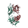 1yecS S: Starting model for refinement |
|---|---|
| Similar structure data |

-Links
 PDBj
PDBj

- Assembly
| Deposited unit | 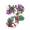
| ||||||||||||
|---|---|---|---|---|---|---|---|---|---|---|---|---|---|
| 1 | 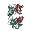
| ||||||||||||
| 2 | 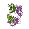
| ||||||||||||
| Unit cell |
| ||||||||||||
| Noncrystallographic symmetry (NCS) | NCS oper:
|
-Components
| #1: Antibody | Mass: 23100.756 Da / Num. of mol.: 2 / Source method: isolated from a natural source / Source: (natural) MUS MUSCULUS (house mouse)#2: Antibody | Mass: 23429.871 Da / Num. of mol.: 2 / Source method: isolated from a natural source / Source: (natural) MUS MUSCULUS (house mouse)#3: Water | ChemComp-HOH / | Water Mass: 18.015 Da / Num. of mol.: 473 / Source method: isolated from a natural source / Formula: H2O Mass: 18.015 Da / Num. of mol.: 473 / Source method: isolated from a natural source / Formula: H2O |
|---|
-Experimental details
-Experiment
| Experiment | Method: X-RAY DIFFRACTION / Number of used crystals: 1 |
|---|
- Sample preparation
| Crystal | Density Matthews: 2.3 Å3/Da / Density % sol: 46 % |
|---|---|
| Crystal grow | pH: 7.5 / Details: 13% PEG8K, 100MM HEPES PH=7.5, 5MM B-OG, pH 7.50 |
-Data collection
| Diffraction | Mean temperature: 77 K |
|---|---|
| Diffraction source | Source: SYNCHROTRON / Site: ESRF  / Beamline: ID29 / Wavelength: 1.08 / Beamline: ID29 / Wavelength: 1.08 |
| Detector | Type: ADSC CCD / Detector: CCD / Date: Jul 15, 2003 |
| Radiation | Protocol: SINGLE WAVELENGTH / Monochromatic (M) / Laue (L): M / Scattering type: x-ray |
| Radiation wavelength | Wavelength: 1.08 Å / Relative weight: 1 |
| Reflection | Resolution: 2→74.536 Å / Num. obs: 60545 / % possible obs: 97 % / Observed criterion σ(I): 3 / Redundancy: 4.9 % / Rmerge(I) obs: 0.091 / Rsym value: 0.091 / Net I/σ(I): 5.9 |
| Reflection shell | Resolution: 2→2.11 Å / Redundancy: 2.8 % / Rmerge(I) obs: 0.291 / Mean I/σ(I) obs: 1.7 / % possible all: 83.9 |
- Processing
| Software |
| ||||||||||||||||||||||||||||||||||||||||||||||||||||||||||||||||||||||||||||||||||||||||||||||||||||||||||||||||||||||||||||||||||||||||||||||||||||||||||||||||||||||||||||||||||||||
|---|---|---|---|---|---|---|---|---|---|---|---|---|---|---|---|---|---|---|---|---|---|---|---|---|---|---|---|---|---|---|---|---|---|---|---|---|---|---|---|---|---|---|---|---|---|---|---|---|---|---|---|---|---|---|---|---|---|---|---|---|---|---|---|---|---|---|---|---|---|---|---|---|---|---|---|---|---|---|---|---|---|---|---|---|---|---|---|---|---|---|---|---|---|---|---|---|---|---|---|---|---|---|---|---|---|---|---|---|---|---|---|---|---|---|---|---|---|---|---|---|---|---|---|---|---|---|---|---|---|---|---|---|---|---|---|---|---|---|---|---|---|---|---|---|---|---|---|---|---|---|---|---|---|---|---|---|---|---|---|---|---|---|---|---|---|---|---|---|---|---|---|---|---|---|---|---|---|---|---|---|---|---|---|
| Refinement | Method to determine structure: MOLECULAR REPLACEMENT Starting model: PDB ENTRY 1YEC Resolution: 2→74.54 Å / Cor.coef. Fo:Fc: 0.942 / Cor.coef. Fo:Fc free: 0.932 / SU B: 3.701 / SU ML: 0.101 / TLS residual ADP flag: LIKELY RESIDUAL / Cross valid method: THROUGHOUT / ESU R: 0.207 / ESU R Free: 0.161 / Stereochemistry target values: MAXIMUM LIKELIHOOD / Details: NONE
| ||||||||||||||||||||||||||||||||||||||||||||||||||||||||||||||||||||||||||||||||||||||||||||||||||||||||||||||||||||||||||||||||||||||||||||||||||||||||||||||||||||||||||||||||||||||
| Solvent computation | Ion probe radii: 0.8 Å / Shrinkage radii: 0.8 Å / VDW probe radii: 1.4 Å / Solvent model: BABINET MODEL WITH MASK | ||||||||||||||||||||||||||||||||||||||||||||||||||||||||||||||||||||||||||||||||||||||||||||||||||||||||||||||||||||||||||||||||||||||||||||||||||||||||||||||||||||||||||||||||||||||
| Displacement parameters | Biso mean: 13.835 Å2
| ||||||||||||||||||||||||||||||||||||||||||||||||||||||||||||||||||||||||||||||||||||||||||||||||||||||||||||||||||||||||||||||||||||||||||||||||||||||||||||||||||||||||||||||||||||||
| Refinement step | Cycle: LAST / Resolution: 2→74.54 Å
| ||||||||||||||||||||||||||||||||||||||||||||||||||||||||||||||||||||||||||||||||||||||||||||||||||||||||||||||||||||||||||||||||||||||||||||||||||||||||||||||||||||||||||||||||||||||
| Refine LS restraints |
|