 Movie
Movie Controller


+ Open data

- Basic information
| Entry | Database: PDB / ID: 1um1 | ||||||
|---|---|---|---|---|---|---|---|
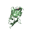
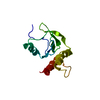
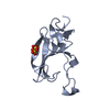
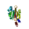
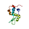
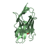
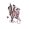
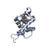
| Title | Solution Structure of RSGI RUH-007, PDZ domain in Human cDNA | ||||||
 Components Components | KIAA1849 protein | ||||||
 Keywords Keywords |  STRUCTURAL GENOMICS / UNKNOWN FUNCTION / PDZ domain / Human cDNA / RIKEN Structural Genomics/Proteomics Initiative / RSGI STRUCTURAL GENOMICS / UNKNOWN FUNCTION / PDZ domain / Human cDNA / RIKEN Structural Genomics/Proteomics Initiative / RSGI | ||||||
| Function / homology |  Function and homology informationneural crest cell migration / substrate adhesion-dependent cell spreading / GTPase binding / microtubule / signal transduction / protein-containing complex Function and homology informationneural crest cell migration / substrate adhesion-dependent cell spreading / GTPase binding / microtubule / signal transduction / protein-containing complexSimilarity search - Function | ||||||
| Biological species |  Homo sapiens (human) Homo sapiens (human) | ||||||
| Method | SOLUTION NMR / torsion angle dynamics | ||||||
 Authors Authors | Doi-Katayama, Y. / Hayashi, F. / Hirota, H. / Yokoyama, S. / RIKEN Structural Genomics/Proteomics Initiative (RSGI) | ||||||
 Citation Citation | Journal: To be Published Title: Solution Structure of RSGI RUH-007, a PDZ Domain in Human cDNA Authors: Doi-Katayama, Y. / Hayashi, F. / Hirota, H. / Yokoyama, S. | ||||||
| History |
|
- Structure visualization
| Structure viewer | Molecule: MolmilJmol/JSmol |
|---|
- Downloads & links
-Download
| PDBx/mmCIF format | 1um1.cif.gz | 620.6 KB | Display | PDBx/mmCIF format |
|---|---|---|---|---|
| PDB format | pdb1um1.ent.gz | 517.3 KB | Display | PDB format |
| PDBx/mmJSON format | 1um1.json.gz | Tree view | PDBx/mmJSON format | |
| Others |  Other downloads Other downloads |
-Validation report
| Arichive directory | https://data.pdbj.org/pub/pdb/validation_reports/um/1um1ftp://data.pdbj.org/pub/pdb/validation_reports/um/1um1 | HTTPS FTP |
|---|
-Related structure data
| Similar structure data | |
|---|---|
| Other databases |
-Links
 PDBj
PDBj

- Assembly
| Deposited unit | 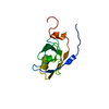
| |||||||||
|---|---|---|---|---|---|---|---|---|---|---|
| 1 |
| |||||||||
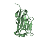
| NMR ensembles |
|
-Components
| #1: Protein | Mass: 11302.925 Da / Num. of mol.: 1 / Fragment: PDZ domain Source method: isolated from a genetically manipulated source Source: (gene. exp.) Homo sapiens (human) / Description: Cell-free protein synthesis / Gene: Kazusa cDNA KIAA1849 / Plasmid: P021030-50 / References: UniProt: Q96JH8 |
|---|
-Experimental details
-Experiment
| Experiment | Method: SOLUTION NMR | ||||||||||||
|---|---|---|---|---|---|---|---|---|---|---|---|---|---|
| NMR experiment |
|
- Sample preparation
| Details | Contents: 0.7mM PDZ domain U-15N, 13C; 20mM Tris-HCl (pH 7.0); 100mM NaCl; 0.02% NaN3; 90% H2O, 10% D2O Solvent system: 90% H2O/10% D2O |
|---|---|
| Sample conditions | Ionic strength: 120mM NaCl / pH: 7.0 / Pressure: ambient / Temperature: 298 K |
-NMR measurement
| Radiation | Protocol: SINGLE WAVELENGTH / Monochromatic (M) / Laue (L): M |
|---|---|
| Radiation wavelength | Relative weight: 1 |
| NMR spectrometer | Type: Varian INOVA / Manufacturer: Varian / Model: INOVA / Field strength: 800 MHz |
- Processing
| NMR software |
| ||||||||||||||||||||||||||||
|---|---|---|---|---|---|---|---|---|---|---|---|---|---|---|---|---|---|---|---|---|---|---|---|---|---|---|---|---|---|
| Refinement | Method: torsion angle dynamics / Software ordinal: 1 | ||||||||||||||||||||||||||||
| NMR representative | Selection criteria: lowest energy | ||||||||||||||||||||||||||||
| NMR ensemble | Conformer selection criteria: structures with the least restraint violations, structures with the lowest energy, target function Conformers calculated total number: 100 / Conformers submitted total number: 20 |
