 Movie
Movie Controller


+ Open data

- Basic information
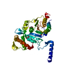
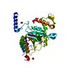
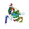
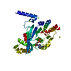
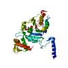
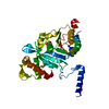
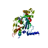
| Entry | Database: PDB / ID: 1ube | ||||||
|---|---|---|---|---|---|---|---|
| Title | MsRecA-ADP Complex | ||||||
 Components Components | RecA | ||||||
 Keywords Keywords | RECOMBINATION / DNA-REPAIR | ||||||
| Function / homology |  Function and homology informationSOS response / ATP-dependent DNA damage sensor activity / single-stranded DNA binding / DNA recombination / damaged DNA binding / DNA repair / ATP hydrolysis activity / ATP binding / cytoplasm Function and homology informationSOS response / ATP-dependent DNA damage sensor activity / single-stranded DNA binding / DNA recombination / damaged DNA binding / DNA repair / ATP hydrolysis activity / ATP binding / cytoplasmSimilarity search - Function | ||||||
| Biological species |  Mycobacterium smegmatis (bacteria) Mycobacterium smegmatis (bacteria) | ||||||
| Method | X-RAY DIFFRACTION / MOLECULAR REPLACEMENT / Resolution: 3.3 Å | ||||||
 Authors Authors | Datta, S. / Krishna, R. / Ganesh, N. / Chandra, N.R. / Muniyappa, K. / Vijayan, M. | ||||||
 Citation Citation | Journal: J.BACTERIOL. / Year: 2003 Title: Crystal Structures of Mycobacterium smegmatis RecA and Its Nucleotide Complexes Authors: Datta, S. / Krishna, R. / Ganesh, N. / Chandra, N.R. / Muniyappa, K. / Vijayan, M. #1: Journal: Nucleic Acids Res. / Year: 2000Title: Crystal Structures of Mycobacterium Tuberculosis Reca and its Complex with Adp-Alf4: Implications For Decreased ATPase Activity and Molecular Aggregation Authors: Datta, S. / Prabu, M. / Vaze, M.B. / Ganesh, N. / Chandra, N.R. / Muniyappa, K. / Vijayan, M. #2: Journal: Proteins / Year: 2003Title: Structural studies on MtRecA-nucleotide complexes: Insights into DNA and nucleotide binding and the structural signature of NTP recognition Authors: Datta, S. / Ganesh, N. / Chandra, N.R. / Muniyappa, K. / Vijayan, M. | ||||||
| History |
|
- Structure visualization
| Structure viewer | Molecule: MolmilJmol/JSmol |
|---|
- Downloads & links
-Download
| PDBx/mmCIF format | 1ube.cif.gz | 67 KB | Display | PDBx/mmCIF format |
|---|---|---|---|---|
| PDB format | pdb1ube.ent.gz | 48.9 KB | Display | PDB format |
| PDBx/mmJSON format | 1ube.json.gz | Tree view | PDBx/mmJSON format | |
| Others |  Other downloads Other downloads |
-Validation report
| Arichive directory | https://data.pdbj.org/pub/pdb/validation_reports/ub/1ubeftp://data.pdbj.org/pub/pdb/validation_reports/ub/1ube | HTTPS FTP |
|---|
-Related structure data
| Related structure data | 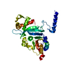 1ubcC 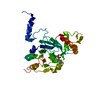 1ubfC 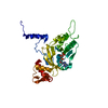 1ubgC 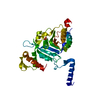 1g19S S: Starting model for refinement C: citing same article ( |
|---|---|
| Similar structure data |


-Links
 PDBj
PDBj
- Assembly
| Deposited unit | 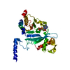
| ||||||||
|---|---|---|---|---|---|---|---|---|---|
| 1 |
| ||||||||
| Unit cell |
|
-Components
| #1: Protein | / Recombinase A Mass: 37344.488 Da / Num. of mol.: 1 Source method: isolated from a genetically manipulated source Source: (gene. exp.) Mycobacterium smegmatis (bacteria) / Plasmid: pThioA / Production host: Escherichia coli (E. coli) / Strain (production host): JC10289 / References: UniProt: Q59560 |
|---|---|
| #2: Chemical | ChemComp-ADP / Adenosine diphosphate  Mass: 427.201 Da / Num. of mol.: 1 / Source method: obtained synthetically / Formula: C10H15N5O10P2 / Comment: ADP, energy-carrying molecule*YM Mass: 427.201 Da / Num. of mol.: 1 / Source method: obtained synthetically / Formula: C10H15N5O10P2 / Comment: ADP, energy-carrying molecule*YM |
| #3: Water | ChemComp-HOH / Water Mass: 18.015 Da / Num. of mol.: 55 / Source method: isolated from a natural source / Formula: H2O Mass: 18.015 Da / Num. of mol.: 55 / Source method: isolated from a natural source / Formula: H2O |
-Experimental details
-Experiment
| Experiment | Method: X-RAY DIFFRACTION / Number of used crystals: 1 |
|---|
- Sample preparation
| Crystal | Density Matthews: 3.5 Å3/Da / Density % sol: 64.57 % | ||||||||||||||||||||||||||||||||||||||||||||||||||||||||
|---|---|---|---|---|---|---|---|---|---|---|---|---|---|---|---|---|---|---|---|---|---|---|---|---|---|---|---|---|---|---|---|---|---|---|---|---|---|---|---|---|---|---|---|---|---|---|---|---|---|---|---|---|---|---|---|---|---|
| Crystal grow | Temperature: 293 K / Method: vapor diffusion, hanging drop / pH: 7.5 Details: PEG 4000, CIT_PHOS, NACL, DTT, pH 7.5, VAPOR DIFFUSION, HANGING DROP, temperature 293K | ||||||||||||||||||||||||||||||||||||||||||||||||||||||||
| Crystal grow | *PLUS Method: vapor diffusion, hanging drop | ||||||||||||||||||||||||||||||||||||||||||||||||||||||||
| Components of the solutions | *PLUS
|
-Data collection
| Diffraction | Mean temperature: 293 K |
|---|---|
| Diffraction source | Source: ROTATING ANODE / Type: RIGAKU RU200 / Wavelength: 1.5418 Å |
| Detector | Type: MARRESEARCH / Detector: IMAGE PLATE / Date: May 14, 2002 |
| Radiation | Monochromator: NULL / Protocol: SINGLE WAVELENGTH / Monochromatic (M) / Laue (L): M / Scattering type: x-ray |
| Radiation wavelength | Wavelength: 1.5418 Å / Relative weight: 1 |
| Reflection | Resolution: 3.3→30 Å / Num. all: 6846 / Num. obs: 6339 / % possible obs: 92.5 % / Observed criterion σ(F): 0 / Observed criterion σ(I): 0 / Redundancy: 2.6 % / Rmerge(I) obs: 0.109 / Net I/σ(I): 5.2 |
| Reflection shell | Resolution: 3.3→3.42 Å / Redundancy: 2.7 % / Rmerge(I) obs: 0.472 / Mean I/σ(I) obs: 1.5 / Num. unique all: 649 / % possible all: 93.8 |
| Reflection | *PLUS Highest resolution: 3.3 Å |
| Reflection shell | *PLUS Highest resolution: 3.3 Å / % possible obs: 93.8 % / Num. unique obs: 649 / Rmerge(I) obs: 0.47 |
- Processing
| Software |
| |||||||||||||||||||||||||
|---|---|---|---|---|---|---|---|---|---|---|---|---|---|---|---|---|---|---|---|---|---|---|---|---|---|---|
| Refinement | Method to determine structure: MOLECULAR REPLACEMENT Starting model: 1g19 Resolution: 3.3→15 Å / Rfactor Rfree error: 0.013 / Data cutoff high absF: 159582.44 / Data cutoff low absF: 0 / Cross valid method: THROUGHOUT / σ(F): 0 / Stereochemistry target values: Engh & Huber
| |||||||||||||||||||||||||
| Solvent computation | Solvent model: FLAT MODEL / Bsol: 51.2327 Å2 / ksol: 0.19912 e/Å3 | |||||||||||||||||||||||||
| Displacement parameters | Biso mean: 65.8 Å2
| |||||||||||||||||||||||||
| Refine analyze |
| |||||||||||||||||||||||||
| Refinement step | Cycle: LAST / Resolution: 3.3→15 Å
| |||||||||||||||||||||||||
| Refine LS restraints |
| |||||||||||||||||||||||||
| LS refinement shell | Resolution: 3.3→3.5 Å / Total num. of bins used: 6
| |||||||||||||||||||||||||
| Xplor file |
| |||||||||||||||||||||||||
| Refinement | *PLUS Highest resolution: 3.3 Å / Lowest resolution: 15 Å / Rfactor Rfree: 0.279 / Rfactor Rwork: 0.228 | |||||||||||||||||||||||||
| Solvent computation | *PLUS | |||||||||||||||||||||||||
| Displacement parameters | *PLUS | |||||||||||||||||||||||||
| Refine LS restraints | *PLUS
|
