 Movie
Movie Controller

[English] 日本語
 Yorodumi
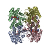
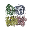
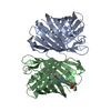
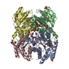
- PDB-1uay: Crystal Structure of Type II 3-Hydroxyacyl-CoA Dehydrogenase from... -
+ Open data

- Basic information
| Entry | Database: PDB / ID: 1uay | ||||||
|---|---|---|---|---|---|---|---|
| Title | Crystal Structure of Type II 3-Hydroxyacyl-CoA Dehydrogenase from Thermus thermophilus HB8 | ||||||
 Components Components | Type II 3-hydroxyacyl-CoA dehydrogenase | ||||||
 Keywords Keywords |  OXIDOREDUCTASE / DEHYDROGENASE / BETA OXIDATION / FATTY ACID / structural genomics / RIKEN Structural Genomics/Proteomics Initiative / RSGI OXIDOREDUCTASE / DEHYDROGENASE / BETA OXIDATION / FATTY ACID / structural genomics / RIKEN Structural Genomics/Proteomics Initiative / RSGI | ||||||
| Function / homology |  Function and homology information Function and homology information | ||||||
| Biological species |   Thermus thermophilus (bacteria) Thermus thermophilus (bacteria) | ||||||
| Method | X-RAY DIFFRACTION / SYNCHROTRON / MOLECULAR REPLACEMENT / Resolution: 1.4 Å | ||||||
 Authors Authors | Kunishima, N. / Asada, Y. / Yokoyama, S. / Kuramitsu, S. / Miyano, M. / RIKEN Structural Genomics/Proteomics Initiative (RSGI) | ||||||
 Citation Citation | Journal: To be Published Title: Crystal Structure of Type II 3-Hydroxyacyl-CoA Dehydrogenase from Thermus thermophilus HB8 Authors: Kunishima, N. / Asada, Y. / Yokoyama, S. / Kuramitsu, S. / Miyano, M. | ||||||
| History |
|
- Structure visualization
| Structure viewer | Molecule: MolmilJmol/JSmol |
|---|
- Downloads & links
-Download
| PDBx/mmCIF format | 1uay.cif.gz | 115.1 KB | Display | PDBx/mmCIF format |
|---|---|---|---|---|
| PDB format | pdb1uay.ent.gz | 87.7 KB | Display | PDB format |
| PDBx/mmJSON format | 1uay.json.gz | Tree view | PDBx/mmJSON format | |
| Others |  Other downloads Other downloads |
-Validation report
| Arichive directory | https://data.pdbj.org/pub/pdb/validation_reports/ua/1uayftp://data.pdbj.org/pub/pdb/validation_reports/ua/1uay | HTTPS FTP |
|---|
-Related structure data
| Related structure data |  1e6wS S: Starting model for refinement |
|---|---|
| Similar structure data | |
| Other databases |


-Links
 PDBj
PDBj


- Assembly
| Deposited unit | 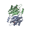
| ||||||||
|---|---|---|---|---|---|---|---|---|---|
| 1 | 
| ||||||||
| Unit cell |
| ||||||||
| Components on special symmetry positions |
| ||||||||
| Details | The biological assembly is tetramer generated from the dimer in the asymmetric unit by the operation: -x+1,y,-z+1/2 |
-Components
| #1: Protein | Mass: 25627.527 Da / Num. of mol.: 2 Source method: isolated from a genetically manipulated source Source: (gene. exp.) Thermus thermophilus (bacteria) / Strain: HB8 / Plasmid: pET11a / Production host: Escherichia coli (E. coli) / References: UniProt: Q7SIA1#2: Chemical | Adenosine  Mass: 267.241 Da / Num. of mol.: 2 / Source method: obtained synthetically / Formula: C10H13N5O4 Mass: 267.241 Da / Num. of mol.: 2 / Source method: obtained synthetically / Formula: C10H13N5O4#3: Water | ChemComp-HOH / | Water Mass: 18.015 Da / Num. of mol.: 584 / Source method: isolated from a natural source / Formula: H2O Mass: 18.015 Da / Num. of mol.: 584 / Source method: isolated from a natural source / Formula: H2O |
|---|
-Experimental details
-Experiment
| Experiment | Method: X-RAY DIFFRACTION / Number of used crystals: 1 |
|---|
- Sample preparation
| Crystal | Density Matthews: 1.79 Å3/Da / Density % sol: 30.69 % |
|---|---|
| Crystal grow | Temperature: 291 K / Method: microbatch / pH: 4.7 Details: sodium phosphate, pH 4.7, MICROBATCH, temperature 291K |
-Data collection
| Diffraction | Mean temperature: 100 K |
|---|---|
| Diffraction source | Source: SYNCHROTRON / Site: SPring-8  / Beamline: BL26B1 / Wavelength: 1 Å / Beamline: BL26B1 / Wavelength: 1 Å |
| Detector | Type: RIGAKU RAXIS V / Detector: IMAGE PLATE / Date: Dec 3, 2002 / Details: mirrors |
| Radiation | Monochromator: Bending Magnet / Protocol: SINGLE WAVELENGTH / Monochromatic (M) / Laue (L): M / Scattering type: x-ray |
| Radiation wavelength | Wavelength: 1 Å / Relative weight: 1 |
| Reflection | Resolution: 1.4→30 Å / Num. all: 83399 / Num. obs: 83399 / % possible obs: 99.2 % / Observed criterion σ(F): 0 / Observed criterion σ(I): 0 / Redundancy: 7.1 % / Biso Wilson estimate: 11.5 Å2 / Rmerge(I) obs: 0.096 / Rsym value: 0.092 / Net I/σ(I): 7.7 |
| Reflection shell | Resolution: 1.4→1.45 Å / Redundancy: 6.81 % / Rmerge(I) obs: 0.75 / Mean I/σ(I) obs: 3.9 / Num. unique all: 8288 / Rsym value: 0.697 / % possible all: 100 |
- Processing
| Software |
| |||||||||||||||||||||||||
|---|---|---|---|---|---|---|---|---|---|---|---|---|---|---|---|---|---|---|---|---|---|---|---|---|---|---|
| Refinement | Method to determine structure: MOLECULAR REPLACEMENT Starting model: PDB ENTRY 1E6W Resolution: 1.4→29.6 Å / Isotropic thermal model: Anisotropic / Cross valid method: THROUGHOUT / σ(F): 0 / Stereochemistry target values: Engh & Huber
| |||||||||||||||||||||||||
| Displacement parameters | Biso mean: 14.1 Å2
| |||||||||||||||||||||||||
| Refine analyze |
| |||||||||||||||||||||||||
| Refinement step | Cycle: LAST / Resolution: 1.4→29.6 Å
| |||||||||||||||||||||||||
| Refine LS restraints |
| |||||||||||||||||||||||||
| LS refinement shell | Resolution: 1.4→1.46 Å / Rfactor Rfree error: 0.01
|
