 Movie
Movie Controller


+ Open data

- Basic information
| Entry | Database: PDB / ID: 1uaa | ||||||
|---|---|---|---|---|---|---|---|
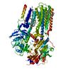
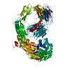
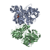
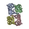
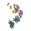
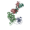
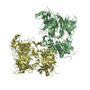
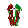
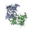
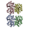
| Title | E. COLI REP HELICASE/DNA COMPLEX | ||||||
 Components Components |
| ||||||
 Keywords Keywords | HYDROLASE/DNA / COMPLEX (HELICASE-DNA) /  HELICASE / DNA UNWINDING / HYDROLASE-DNA COMPLEX HELICASE / DNA UNWINDING / HYDROLASE-DNA COMPLEX | ||||||
| Function / homology |  Function and homology information Function and homology informationbacterial-type DNA replication / recombinational repair / 3'-5' DNA helicase activity / DNA unwinding involved in DNA replication / 3'-5' exonuclease activity / DNA helicase activity / response to radiation / single-stranded DNA binding / DNA helicase / DNA repair ...bacterial-type DNA replication / recombinational repair / 3'-5' DNA helicase activity / DNA unwinding involved in DNA replication / 3'-5' exonuclease activity / DNA helicase activity / response to radiation / single-stranded DNA binding / DNA helicase / DNA repair / ATP binding / cytosolSimilarity search - Function | ||||||
| Biological species |  Escherichia coli (E. coli) Escherichia coli (E. coli) | ||||||
| Method | X-RAY DIFFRACTION / SYNCHROTRON / Resolution: 3 Å | ||||||
 Authors Authors | Korolev, S. / Waksman, G. | ||||||
 Citation Citation | Journal: Cell(Cambridge,Mass.) / Year: 1997 Title: Major domain swiveling revealed by the crystal structures of complexes of E. coli Rep helicase bound to single-stranded DNA and ADP. Authors: Korolev, S. / Hsieh, J. / Gauss, G.H. / Lohman, T.M. / Waksman, G. | ||||||
| History |
|
- Structure visualization
| Structure viewer | Molecule: MolmilJmol/JSmol |
|---|
- Downloads & links
-Download
| PDBx/mmCIF format | 1uaa.cif.gz | 262.2 KB | Display | PDBx/mmCIF format |
|---|---|---|---|---|
| PDB format | pdb1uaa.ent.gz | 212.7 KB | Display | PDB format |
| PDBx/mmJSON format | 1uaa.json.gz | Tree view | PDBx/mmJSON format | |
| Others |  Other downloads Other downloads |
-Validation report
| Arichive directory | https://data.pdbj.org/pub/pdb/validation_reports/ua/1uaaftp://data.pdbj.org/pub/pdb/validation_reports/ua/1uaa | HTTPS FTP |
|---|
-Related structure data
| Similar structure data |
|---|
-Links
 PDBj
PDBj


- Assembly
| Deposited unit | 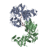
| ||||||||
|---|---|---|---|---|---|---|---|---|---|
| 1 |
| ||||||||
| Unit cell |
|
-Components
| #1: DNA chain | Mass: 4822.127 Da / Num. of mol.: 1 Source method: isolated from a genetically manipulated source Source: (gene. exp.) Escherichia coli (E. coli) |
|---|---|
| #2: Protein | Mass: 77042.078 Da / Num. of mol.: 2 / Source method: obtained synthetically References: UniProt: P09980, Hydrolases; Acting on acid anhydrides; In phosphorus-containing anhydrides |
-Experimental details
-Experiment
| Experiment | Method: X-RAY DIFFRACTION |
|---|
- Sample preparation
| Crystal | Density Matthews: 4.5 Å3/Da / Density % sol: 70 % | ||||||||||||||||||||||||||||||||||||||||||||||||
|---|---|---|---|---|---|---|---|---|---|---|---|---|---|---|---|---|---|---|---|---|---|---|---|---|---|---|---|---|---|---|---|---|---|---|---|---|---|---|---|---|---|---|---|---|---|---|---|---|---|
| Crystal grow | Temperature: 293 K / Method: vapor diffusion / pH: 8 / Details: pH 8.00, VAPOR DIFFUSION, temperature 293.00K | ||||||||||||||||||||||||||||||||||||||||||||||||
| Components of the solutions |
| ||||||||||||||||||||||||||||||||||||||||||||||||
| Crystal | *PLUS | ||||||||||||||||||||||||||||||||||||||||||||||||
| Crystal grow | *PLUS Temperature: 20 ℃ / pH: 8 / Method: vapor diffusion, hanging drop | ||||||||||||||||||||||||||||||||||||||||||||||||
| Components of the solutions | *PLUS
|
-Data collection
| Diffraction | Mean temperature: 100 K |
|---|---|
| Diffraction source | Source: SYNCHROTRON / Site: SSRL  / Beamline: BL7-1 / Beamline: BL7-1 |
| Detector | Type: MARRESEARCH / Detector: IMAGE PLATE / Date: Apr 7, 1996 |
| Radiation | Protocol: SINGLE WAVELENGTH / Monochromatic (M) / Laue (L): M / Scattering type: x-ray |
| Radiation wavelength | Relative weight: 1 |
| Reflection | Resolution: 3→30 Å / Num. obs: 57472 / % possible obs: 97.3 % / Redundancy: 7 % / Rmerge(I) obs: 0.053 |
| Reflection | *PLUS Highest resolution: 3 Å / Lowest resolution: 30 Å / % possible obs: 97.3 % / Num. measured all: 434546 |
- Processing
| Software |
| ||||||||||||||||||||||||||||||||||||||||||||||||||||||||||||
|---|---|---|---|---|---|---|---|---|---|---|---|---|---|---|---|---|---|---|---|---|---|---|---|---|---|---|---|---|---|---|---|---|---|---|---|---|---|---|---|---|---|---|---|---|---|---|---|---|---|---|---|---|---|---|---|---|---|---|---|---|---|
| Refinement | Resolution: 3→15 Å / σ(F): 2
| ||||||||||||||||||||||||||||||||||||||||||||||||||||||||||||
| Displacement parameters | Biso mean: 41 Å2 | ||||||||||||||||||||||||||||||||||||||||||||||||||||||||||||
| Refinement step | Cycle: LAST / Resolution: 3→15 Å
| ||||||||||||||||||||||||||||||||||||||||||||||||||||||||||||
| Refine LS restraints |
| ||||||||||||||||||||||||||||||||||||||||||||||||||||||||||||
| Software | *PLUS Name: X-PLOR / Version: 3.8 / Classification: refinement | ||||||||||||||||||||||||||||||||||||||||||||||||||||||||||||
| Refinement | *PLUS Highest resolution: 3 Å / Lowest resolution: 15 Å / σ(I): 2 / Rfactor all: 0.235 / Rfactor obs: 0.228 | ||||||||||||||||||||||||||||||||||||||||||||||||||||||||||||
| Solvent computation | *PLUS | ||||||||||||||||||||||||||||||||||||||||||||||||||||||||||||
| Displacement parameters | *PLUS Biso mean: 41 Å2 | ||||||||||||||||||||||||||||||||||||||||||||||||||||||||||||
| Refine LS restraints | *PLUS Type: x_angle_deg / Dev ideal: 1.918 |
