 Movie
Movie Controller

[English] 日本語
 Yorodumi
- PDB-1u7r: Crystal structure of Native Sperm Whale myoglobin from low ionic ... -
+ Open data

- Basic information
| Entry | Database: PDB / ID: 1u7r | ||||||
|---|---|---|---|---|---|---|---|
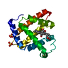
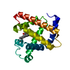
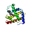
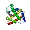
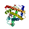
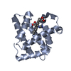
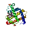
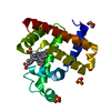
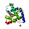
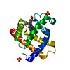
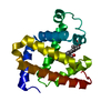
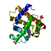
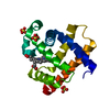
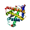
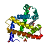
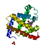
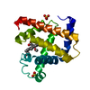
| Title | Crystal structure of Native Sperm Whale myoglobin from low ionic strength enviroment (Form2 ) | ||||||
 Components Components | Myoglobin | ||||||
 Keywords Keywords | TRANSPORT PROTEIN / Sperm Whale myoglobin / Low ionic strength | ||||||
| Function / homology |  Function and homology information Function and homology informationhydrogen peroxide mediated signaling pathway / oxygen carrier activity / oxygen binding / heme binding / metal ion bindingSimilarity search - Function | ||||||
| Biological species |  Physeter catodon (sperm whale) Physeter catodon (sperm whale) | ||||||
| Method | X-RAY DIFFRACTION / SYNCHROTRON / MOLECULAR REPLACEMENT / Resolution: 1.15 Å | ||||||
 Authors Authors | Zhang, W. / Phillips Jr., G.N. | ||||||
 Citation Citation | Journal: Proteins / Year: 2008 Title: Sampling of the native conformational ensemble of myoglobin via structures in different crystalline environments. Authors: Kondrashov, D.A. / Zhang, W. / Aranda, R. / Stec, B. / Phillips Jr., G.N. | ||||||
| History |
|
- Structure visualization
| Structure viewer | Molecule: MolmilJmol/JSmol |
|---|
- Downloads & links
-Download
| PDBx/mmCIF format | 1u7r.cif.gz | 84.3 KB | Display | PDBx/mmCIF format |
|---|---|---|---|---|
| PDB format | pdb1u7r.ent.gz | 62.9 KB | Display | PDB format |
| PDBx/mmJSON format | 1u7r.json.gz | Tree view | PDBx/mmJSON format | |
| Others |  Other downloads Other downloads |
-Validation report
| Arichive directory | https://data.pdbj.org/pub/pdb/validation_reports/u7/1u7rftp://data.pdbj.org/pub/pdb/validation_reports/u7/1u7r | HTTPS FTP |
|---|
-Related structure data


-Links
 PDBj
PDBj


- Assembly
| Deposited unit | 
| ||||||||
|---|---|---|---|---|---|---|---|---|---|
| 1 |
| ||||||||
| Unit cell |
|
-Components
| #1: Protein | Mass: 17234.951 Da / Num. of mol.: 1 / Source method: isolated from a natural source / Source: (natural) Physeter catodon (sperm whale) / Tissue: skeleton / References: UniProt: P02185 |
|---|---|
| #2: Chemical | ChemComp-HEM / Heme B  Mass: 616.487 Da / Num. of mol.: 1 / Source method: obtained synthetically / Formula: C34H32FeN4O4 Mass: 616.487 Da / Num. of mol.: 1 / Source method: obtained synthetically / Formula: C34H32FeN4O4 |
| #3: Chemical | ChemComp-IMD / Imidazole  Mass: 69.085 Da / Num. of mol.: 1 / Source method: obtained synthetically / Formula: C3H5N2 Mass: 69.085 Da / Num. of mol.: 1 / Source method: obtained synthetically / Formula: C3H5N2 |
| #4: Water | ChemComp-HOH / Water Mass: 18.015 Da / Num. of mol.: 183 / Source method: isolated from a natural source / Formula: H2O Mass: 18.015 Da / Num. of mol.: 183 / Source method: isolated from a natural source / Formula: H2O |
-Experimental details
-Experiment
| Experiment | Method: X-RAY DIFFRACTION / Number of used crystals: 1 |
|---|
- Sample preparation
| Crystal | Density Matthews: 1.83 Å3/Da / Density % sol: 32.8 % |
|---|---|
| Crystal grow | Temperature: 277 K / Method: vapor diffusion, hanging drop / pH: 7 Details: 50mM imidazole, pH 7 with 16% PEG3550, VAPOR DIFFUSION, HANGING DROP, temperature 277K |
-Data collection
| Diffraction | Mean temperature: 100 K |
|---|---|
| Diffraction source | Source: SYNCHROTRON / Site: APS  / Beamline: 14-BM-D / Wavelength: 1 Å / Beamline: 14-BM-D / Wavelength: 1 Å |
| Detector | Type: ADSC QUANTUM 4 / Detector: CCD / Date: Jan 1, 2000 |
| Radiation | Monochromator: GRAPHITE / Protocol: SINGLE WAVELENGTH / Monochromatic (M) / Laue (L): M / Scattering type: x-ray |
| Radiation wavelength | Wavelength: 1 Å / Relative weight: 1 |
| Reflection | Resolution: 1.15→40 Å / Num. all: 53703 / Num. obs: 53703 / % possible obs: 100 % / Observed criterion σ(F): 0 / Observed criterion σ(I): -1000 / Rmerge(I) obs: 0.039 |
| Reflection shell | Resolution: 1.15→1.19 Å / Rmerge(I) obs: 0.235 / Mean I/σ(I) obs: 2.8 / Rsym value: 0.039 / % possible all: 92.8 |
- Processing
| Software |
| |||||||||||||||||||||||||||||||||
|---|---|---|---|---|---|---|---|---|---|---|---|---|---|---|---|---|---|---|---|---|---|---|---|---|---|---|---|---|---|---|---|---|---|---|
| Refinement | Method to determine structure: MOLECULAR REPLACEMENT / Resolution: 1.15→40 Å / Num. parameters: 13055 / Num. restraintsaints: 16112 / Isotropic thermal model: Anisotropic / Cross valid method: FREE R / σ(F): 0 / Stereochemistry target values: ENGH AND HUBER / Details: ANISOTROPIC REFINEMENT REDUCED FREE R (NO CUTOFF)
| |||||||||||||||||||||||||||||||||
| Refine analyze | Num. disordered residues: 5 / Occupancy sum hydrogen: 0 / Occupancy sum non hydrogen: 1413 | |||||||||||||||||||||||||||||||||
| Refinement step | Cycle: LAST / Resolution: 1.15→40 Å
| |||||||||||||||||||||||||||||||||
| Refine LS restraints |
| |||||||||||||||||||||||||||||||||
| LS refinement shell | Resolution: 1.15→1.19 Å
|
