 Movie
Movie Controller

[English] 日本語
 Yorodumi
- PDB-1u25: Crystal structure of Selenomonas ruminantium phytase complexed wi... -
+ Open data

- Basic information
| Entry | Database: PDB / ID: 1u25 | ||||||
|---|---|---|---|---|---|---|---|
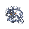
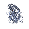
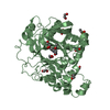
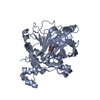
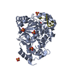
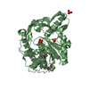
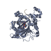
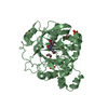
| Title | Crystal structure of Selenomonas ruminantium phytase complexed with persulfated phytate in the C2221 crystal form | ||||||
 Components Components | myo-inositol hexaphosphate phosphohydrolase | ||||||
 Keywords Keywords |  HYDROLASE / PTP / P-loop / phytase HYDROLASE / PTP / P-loop / phytase | ||||||
| Function / homology |  Function and homology information Function and homology information | ||||||
| Biological species |  Selenomonas ruminantium (bacteria) Selenomonas ruminantium (bacteria) | ||||||
| Method | X-RAY DIFFRACTION / SYNCHROTRON / MOLECULAR REPLACEMENT / Resolution: 2.5 Å | ||||||
 Authors Authors | Chu, H.M. / Guo, R.T. / Lin, T.W. / Chou, C.C. / Shr, H.L. / Lai, H.L. / Tang, T.Y. / Cheng, K.J. / Selinger, B.L. / Wang, A.H.-J. | ||||||
 Citation Citation | Journal: STRUCTURE / Year: 2004 Title: Structures of Selenomonas ruminantium Phytase in Complex with Persulfated Phytate; DSP Phytase Fold and Mechanism for Sequential Substrate Hydrolysis Authors: Chu, H.M. / Guo, R.T. / Lin, T.W. / Chou, C.C. / Shr, H.L. / Lai, H.L. / Tang, T.Y. / Cheng, K.J. / Selinger, B.L. / Wang, A.H.-J. | ||||||
| History |
|
- Structure visualization
| Structure viewer | Molecule: MolmilJmol/JSmol |
|---|
- Downloads & links
-Download
| PDBx/mmCIF format | 1u25.cif.gz | 218 KB | Display | PDBx/mmCIF format |
|---|---|---|---|---|
| PDB format | pdb1u25.ent.gz | 184 KB | Display | PDB format |
| PDBx/mmJSON format | 1u25.json.gz | Tree view | PDBx/mmJSON format | |
| Others |  Other downloads Other downloads |
-Validation report
| Arichive directory | https://data.pdbj.org/pub/pdb/validation_reports/u2/1u25ftp://data.pdbj.org/pub/pdb/validation_reports/u2/1u25 | HTTPS FTP |
|---|
-Related structure data


-Links
 PDBj
PDBj


- Assembly
| Deposited unit | 
| ||||||||||||||||||||||||||||||
|---|---|---|---|---|---|---|---|---|---|---|---|---|---|---|---|---|---|---|---|---|---|---|---|---|---|---|---|---|---|---|---|
| 1 | 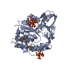
| ||||||||||||||||||||||||||||||
| 2 | 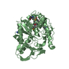
| ||||||||||||||||||||||||||||||
| 3 | 
| ||||||||||||||||||||||||||||||
| Unit cell |
| ||||||||||||||||||||||||||||||
| Components on special symmetry positions |
|
-Components
| #1: Protein | Mass: 39007.547 Da / Num. of mol.: 3 Source method: isolated from a genetically manipulated source Source: (gene. exp.) Selenomonas ruminantium (bacteria) / Plasmid: pET23b / Production host: Escherichia coli (E. coli) / Strain (production host): BL21 / References: UniProt: Q7WUJ1, EC: 3.1.3.72#2: Chemical |   Mass: 660.535 Da / Num. of mol.: 3 / Source method: obtained synthetically / Formula: C6H12O24S6 Mass: 660.535 Da / Num. of mol.: 3 / Source method: obtained synthetically / Formula: C6H12O24S6#3: Water | ChemComp-HOH / | Water Mass: 18.015 Da / Num. of mol.: 941 / Source method: isolated from a natural source / Formula: H2O Mass: 18.015 Da / Num. of mol.: 941 / Source method: isolated from a natural source / Formula: H2O |
|---|
-Experimental details
-Experiment
| Experiment | Method: X-RAY DIFFRACTION / Number of used crystals: 1 |
|---|
- Sample preparation
| Crystal | Density Matthews: 3.035 Å3/Da / Density % sol: 57.91 % |
|---|---|
| Crystal grow | Temperature: 298 K / Method: vapor diffusion, hanging drop / pH: 9 Details: 0.1M LiCl, 0.05M CAPSO, 12% PEG6000, 2% PEG200, pH 9.0, VAPOR DIFFUSION, HANGING DROP, temperature 298K |
-Data collection
| Diffraction source | Source: SYNCHROTRON / Site: SPring-8  / Beamline: BL12B2 / Wavelength: 1.0362 Å / Beamline: BL12B2 / Wavelength: 1.0362 Å |
|---|---|
| Detector | Type: ADSC QUANTUM 4 / Detector: CCD / Date: Feb 15, 2003 / Details: mirror |
| Radiation | Protocol: SINGLE WAVELENGTH / Monochromatic (M) / Laue (L): M / Scattering type: x-ray |
| Radiation wavelength | Wavelength: 1.0362 Å / Relative weight: 1 |
| Reflection | Resolution: 2.44→50 Å / Num. all: 47108 / Num. obs: 37388 / % possible obs: 84 % / Observed criterion σ(F): 2 / Observed criterion σ(I): 2 / Rmerge(I) obs: 0.078 / Net I/σ(I): 13 |
| Reflection shell | Resolution: 2.44→2.53 Å |
- Processing
| Software |
| ||||||||||||||||||||
|---|---|---|---|---|---|---|---|---|---|---|---|---|---|---|---|---|---|---|---|---|---|
| Refinement | Method to determine structure: MOLECULAR REPLACEMENT / Resolution: 2.5→35.21 Å / σ(F): 2 / Stereochemistry target values: Engh & Huber
| ||||||||||||||||||||
| Refine analyze |
| ||||||||||||||||||||
| Refinement step | Cycle: LAST / Resolution: 2.5→35.21 Å
| ||||||||||||||||||||
| Refine LS restraints |
| ||||||||||||||||||||
| LS refinement shell | Resolution: 2.5→2.66 Å / Rfactor Rfree error: 0.024
|
