 Movie
Movie Controller


+ Open data

- Basic information
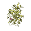
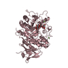
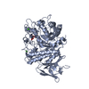
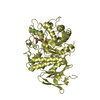
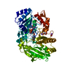
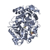
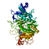
| Entry | Database: PDB / ID: 1txn | ||||||
|---|---|---|---|---|---|---|---|
| Title | Crystal structure of coproporphyrinogen III oxidase | ||||||
 Components Components | Coproporphyrinogen III oxidase | ||||||
 Keywords Keywords | OXIDOREDUCTASE / Structural genomics / Dimer / Novel fold / PSI / Protein Structure Initiative / New York SGX Research Center for Structural Genomics / NYSGXRC | ||||||
| Function / homology |  Function and homology informationcoproporphyrinogen oxidase / coproporphyrinogen oxidase activity / Heme biosynthesis / protoporphyrinogen IX biosynthetic process / heme biosynthetic process / protein homodimerization activity / cytosol / cytoplasm Function and homology informationcoproporphyrinogen oxidase / coproporphyrinogen oxidase activity / Heme biosynthesis / protoporphyrinogen IX biosynthetic process / heme biosynthetic process / protein homodimerization activity / cytosol / cytoplasmSimilarity search - Function | ||||||
| Biological species |  Saccharomyces cerevisiae (brewer's yeast) Saccharomyces cerevisiae (brewer's yeast) | ||||||
| Method | X-RAY DIFFRACTION / SYNCHROTRON / SAD / Resolution: 1.7 Å | ||||||
 Authors Authors | Kumaran, D. / Swaminathan, S. / Burley, S.K. / New York SGX Research Center for Structural Genomics (NYSGXRC) | ||||||
 Citation Citation | Journal: To be Published Title: Crystal structure of coproporphyrinogen III oxidase Authors: Kumaran, D. / Swaminathan, S. | ||||||
| History |
|
- Structure visualization
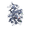
| Structure viewer | Molecule: MolmilJmol/JSmol |
|---|
- Downloads & links
-Download
| PDBx/mmCIF format | 1txn.cif.gz | 122.5 KB | Display | PDBx/mmCIF format |
|---|---|---|---|---|
| PDB format | pdb1txn.ent.gz | 100.3 KB | Display | PDB format |
| PDBx/mmJSON format | 1txn.json.gz | Tree view | PDBx/mmJSON format | |
| Others |  Other downloads Other downloads |
-Validation report
| Arichive directory | https://data.pdbj.org/pub/pdb/validation_reports/tx/1txnftp://data.pdbj.org/pub/pdb/validation_reports/tx/1txn | HTTPS FTP |
|---|
-Related structure data
| Similar structure data | |
|---|---|
| Other databases |

-Links
 PDBj
PDBj
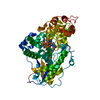
- Assembly
| Deposited unit | 
| ||||||||
|---|---|---|---|---|---|---|---|---|---|
| 1 |
| ||||||||
| Unit cell |
|
-Components
| #1: Protein | / Coproporphyrinogenase / Coprogen oxidase / COX Mass: 38138.699 Da / Num. of mol.: 2 Source method: isolated from a genetically manipulated source Source: (gene. exp.) Saccharomyces cerevisiae (brewer's yeast)Gene: HEM13, YDR044W, YD5112.02 / Production host:  Escherichia coli (E. coli) / References: UniProt: P11353, coproporphyrinogen oxidase Escherichia coli (E. coli) / References: UniProt: P11353, coproporphyrinogen oxidase#2: Chemical | ChemComp-GOL / | Glycerol  Mass: 92.094 Da / Num. of mol.: 1 / Source method: obtained synthetically / Formula: C3H8O3 Mass: 92.094 Da / Num. of mol.: 1 / Source method: obtained synthetically / Formula: C3H8O3#3: Water | ChemComp-HOH / | Water Mass: 18.015 Da / Num. of mol.: 431 / Source method: isolated from a natural source / Formula: H2O Mass: 18.015 Da / Num. of mol.: 431 / Source method: isolated from a natural source / Formula: H2O |
|---|
-Experimental details
-Experiment
| Experiment | Method: X-RAY DIFFRACTION / Number of used crystals: 1 |
|---|
- Sample preparation
| Crystal | Density Matthews: 2 Å3/Da / Density % sol: 40 % |
|---|---|
| Crystal grow | Temperature: 293 K / Method: vapor diffusion, sitting drop / pH: 6.5 Details: PEG 8000, Sodium Cacodylate, Magnesium acetate, pH 6.5, VAPOR DIFFUSION, SITTING DROP, temperature 293K |
-Data collection
| Diffraction | Mean temperature: 100 K |
|---|---|
| Diffraction source | Source: SYNCHROTRON / Site: NSLS  / Beamline: X12C / Wavelength: 0.979 Å / Beamline: X12C / Wavelength: 0.979 Å |
| Detector | Type: BRANDEIS - B4 / Detector: CCD / Date: Feb 6, 2003 / Details: mirrors |
| Radiation | Monochromator: Si 111 / Protocol: SINGLE WAVELENGTH / Monochromatic (M) / Laue (L): M / Scattering type: x-ray |
| Radiation wavelength | Wavelength: 0.979 Å / Relative weight: 1 |
| Reflection | Resolution: 1.7→50 Å / Num. all: 61551 / Num. obs: 61551 / % possible obs: 94.2 % / Observed criterion σ(F): 0 / Observed criterion σ(I): 0 / Redundancy: 13.2 % / Rmerge(I) obs: 0.081 / Net I/σ(I): 15.5 |
| Reflection shell | Resolution: 1.7→1.75 Å / Rmerge(I) obs: 0.379 / Num. unique all: 3677 / % possible all: 87.4 |
- Processing
| Software |
| |||||||||||||||||||||||||
|---|---|---|---|---|---|---|---|---|---|---|---|---|---|---|---|---|---|---|---|---|---|---|---|---|---|---|
| Refinement | Method to determine structure: SAD / Resolution: 1.7→50 Å / Isotropic thermal model: Isotropic / Cross valid method: THROUGHOUT / σ(F): 0 / Stereochemistry target values: Engh & Huber Details: Residues 97-106 and 271-328 in chain A, and residues 97-106, 203-205, and 271-328 in chain B, were not visbile in the electron density and hence not modeled
| |||||||||||||||||||||||||
| Displacement parameters | Biso mean: 34.5 Å2 | |||||||||||||||||||||||||
| Refinement step | Cycle: LAST / Resolution: 1.7→50 Å
| |||||||||||||||||||||||||
| Refine LS restraints |
| |||||||||||||||||||||||||
| LS refinement shell | Resolution: 1.7→1.71 Å /
|
