 Movie
Movie Controller


+ Open data

- Basic information
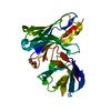
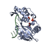
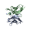
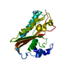
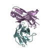
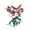
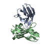
| Entry | Database: PDB / ID: 1tvd | ||||||
|---|---|---|---|---|---|---|---|
| Title | VARIABLE DOMAIN OF T CELL RECEPTOR DELTA CHAIN | ||||||
 Components Components | T CELL RECEPTOR T-cell receptor T-cell receptor | ||||||
 Keywords Keywords | IMMUNORECEPTOR / TCR / DELTA CHAIN / VARIABLE DOMAIN | ||||||
| Function / homology |  Function and homology informationT cell receptor complex / adaptive immune response / immune response / innate immune response / extracellular space Function and homology informationT cell receptor complex / adaptive immune response / immune response / innate immune response / extracellular spaceSimilarity search - Function | ||||||
| Biological species |  Homo sapiens (human) Homo sapiens (human) | ||||||
| Method | X-RAY DIFFRACTION / SYNCHROTRON / MIRAS / Resolution: 1.9 Å | ||||||
 Authors Authors | Li, H.M. / Lebedeva, M.I. / Llera, A.S. / Fields, B.A. / Brenner, M.B. / Mariuzza, R.A. | ||||||
 Citation Citation | Journal: Nature / Year: 1998 Title: Structure of the Vdelta domain of a human gammadelta T-cell antigen receptor. Authors: Li, H. / Lebedeva, M.I. / Llera, A.S. / Fields, B.A. / Brenner, M.B. / Mariuzza, R.A. #1: Journal: Protein Sci. / Year: 1996Title: Cloning, Expression, and Crystallization of the V Delta Domain of a Human Gamma Delta T-Cell Receptor Authors: Lebedeva, M.I. / Fields, B.A. / Spits, H. / Panchamoorthy, G. / Brenner, M.B. / Mariuzza, R.A. | ||||||
| History |
|
- Structure visualization
| Structure viewer | Molecule: MolmilJmol/JSmol |
|---|
- Downloads & links
-Download
| PDBx/mmCIF format | 1tvd.cif.gz | 61.7 KB | Display | PDBx/mmCIF format |
|---|---|---|---|---|
| PDB format | pdb1tvd.ent.gz | 49.5 KB | Display | PDB format |
| PDBx/mmJSON format | 1tvd.json.gz | Tree view | PDBx/mmJSON format | |
| Others |  Other downloads Other downloads |
-Validation report
| Arichive directory | https://data.pdbj.org/pub/pdb/validation_reports/tv/1tvdftp://data.pdbj.org/pub/pdb/validation_reports/tv/1tvd | HTTPS FTP |
|---|
-Related structure data
| Similar structure data |
|---|


-Links
 PDBj
PDBj- Assembly
| Deposited unit | 
| ||||||||
|---|---|---|---|---|---|---|---|---|---|
| 1 |
| ||||||||
| Unit cell |
| ||||||||
| Noncrystallographic symmetry (NCS) | NCS oper: (Code: given Matrix: (-0.971829, -0.032294, -0.233465), Vector : |
-Components
| #1: Protein | T-cell receptor / ES204 V DELTA Mass: 13145.624 Da / Num. of mol.: 2 / Fragment: DELTA CHAIN, VARIABLE DOMAIN Source method: isolated from a genetically manipulated source Source: (gene. exp.) Homo sapiens (human) / Cell: T-LYMPHOCYTE / Cell line: ES204 / Plasmid: PET26B(+) / Cellular location (production host): PERIPLASM / Production host:  Escherichia coli (E. coli) / Strain (production host): K-12 BL21 (DE3) / References: PIR: S04934, UniProt: A0JD37*PLUS Escherichia coli (E. coli) / Strain (production host): K-12 BL21 (DE3) / References: PIR: S04934, UniProt: A0JD37*PLUS#2: Water | ChemComp-HOH / | Water Mass: 18.015 Da / Num. of mol.: 309 / Source method: isolated from a natural source / Formula: H2O Mass: 18.015 Da / Num. of mol.: 309 / Source method: isolated from a natural source / Formula: H2O |
|---|
-Experimental details
-Experiment
| Experiment | Method: X-RAY DIFFRACTION / Number of used crystals: 2 |
|---|
- Sample preparation
| Crystal | Density Matthews: 2.4 Å3/Da / Density % sol: 40 % | ||||||||||||||||||||||||
|---|---|---|---|---|---|---|---|---|---|---|---|---|---|---|---|---|---|---|---|---|---|---|---|---|---|
| Crystal grow | pH: 6.5 Details: 0.65-0.75M SODIUM CITRATE 0.1M SODIUM CACODYLATE, PH 6.5 | ||||||||||||||||||||||||
| Crystal | *PLUS | ||||||||||||||||||||||||
| Crystal grow | *PLUS Method: vapor diffusion, hanging drop / Details: Lebedeva, M.I., (1996) Protein Sci., 5, 2638. / pH: 7 | ||||||||||||||||||||||||
| Components of the solutions | *PLUS
|
-Data collection
| Diffraction | Mean temperature: 278 K |
|---|---|
| Diffraction source | Source: SYNCHROTRON / Site: NSLS  / Beamline: X8C / Wavelength: 1 / Beamline: X8C / Wavelength: 1 |
| Detector | Type: SIEMENS / Detector: CCD / Date: Jun 1, 1996 |
| Radiation | Monochromatic (M) / Laue (L): M / Scattering type: x-ray |
| Radiation wavelength | Wavelength: 1 Å / Relative weight: 1 |
| Reflection | Resolution: 1.9→99 Å / Num. obs: 44342 / % possible obs: 83.3 % / Redundancy: 3 % / Rmerge(I) obs: 0.093 / Rsym value: 0.093 / Net I/σ(I): 10 |
| Reflection shell | Resolution: 1.9→2 Å / Redundancy: 2 % / Rmerge(I) obs: 0.166 / Mean I/σ(I) obs: 3 / Rsym value: 0.166 / % possible all: 53.7 |
| Reflection | *PLUS Num. obs: 13905 / Num. measured all: 44342 |
- Processing
| Software |
| ||||||||||||||||||||||||||||||||||||||||||||||||||||||||||||
|---|---|---|---|---|---|---|---|---|---|---|---|---|---|---|---|---|---|---|---|---|---|---|---|---|---|---|---|---|---|---|---|---|---|---|---|---|---|---|---|---|---|---|---|---|---|---|---|---|---|---|---|---|---|---|---|---|---|---|---|---|---|
| Refinement | Method to determine structure: MIRAS / Resolution: 1.9→6 Å / Cross valid method: THROUGHOUT / σ(F): 0
| ||||||||||||||||||||||||||||||||||||||||||||||||||||||||||||
| Displacement parameters | Biso mean: 14.8 Å2 | ||||||||||||||||||||||||||||||||||||||||||||||||||||||||||||
| Refinement step | Cycle: LAST / Resolution: 1.9→6 Å
| ||||||||||||||||||||||||||||||||||||||||||||||||||||||||||||
| Refine LS restraints |
| ||||||||||||||||||||||||||||||||||||||||||||||||||||||||||||
| LS refinement shell | Resolution: 1.9→1.99 Å / Total num. of bins used: 15
| ||||||||||||||||||||||||||||||||||||||||||||||||||||||||||||
| Xplor file |
| ||||||||||||||||||||||||||||||||||||||||||||||||||||||||||||
| Software | *PLUS Name: X-PLOR / Version: 3.1 / Classification: refinement | ||||||||||||||||||||||||||||||||||||||||||||||||||||||||||||
| Refinement | *PLUS Rfactor all: 0.174 | ||||||||||||||||||||||||||||||||||||||||||||||||||||||||||||
| Solvent computation | *PLUS | ||||||||||||||||||||||||||||||||||||||||||||||||||||||||||||
| Displacement parameters | *PLUS | ||||||||||||||||||||||||||||||||||||||||||||||||||||||||||||
| Refine LS restraints | *PLUS
|
