 Movie
Movie Controller


+ Open data

- Basic information
| Entry | Database: PDB / ID: 1tr9 | ||||||
|---|---|---|---|---|---|---|---|
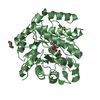
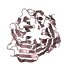
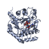
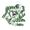
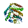
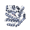
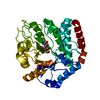
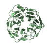
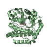
| Title | Structure of beta-hexosaminidase from Vibrio cholerae | ||||||
 Components Components | Beta-hexosaminidase Hexosaminidase Hexosaminidase | ||||||
 Keywords Keywords | HYDROLASE / beta-alpha barrel / Beta-hexosaminidase / Structural Genomics / PSI / Protein Structure Initiative / New York SGX Research Center for Structural Genomics / NYSGXRC | ||||||
| Function / homology |  Function and homology information Function and homology informationpeptidoglycan turnover / beta-N-acetylhexosaminidase / beta-N-acetylhexosaminidase activity / N-acetyl-beta-D-galactosaminidase activity / peptidoglycan biosynthetic process / cell wall organization / regulation of cell shape / carbohydrate metabolic process / cell cycle / cell division ...peptidoglycan turnover / beta-N-acetylhexosaminidase / beta-N-acetylhexosaminidase activity / N-acetyl-beta-D-galactosaminidase activity / peptidoglycan biosynthetic process / cell wall organization / regulation of cell shape / carbohydrate metabolic process / cell cycle / cell division / response to antibiotic / cytoplasmSimilarity search - Function | ||||||
| Biological species |   Vibrio cholerae (bacteria) Vibrio cholerae (bacteria) | ||||||
| Method | X-RAY DIFFRACTION / SYNCHROTRON / SAD / Resolution: 1.8 Å | ||||||
 Authors Authors | Gorman, J. / Shapiro, L. / Burley, S.K. / New York SGX Research Center for Structural Genomics (NYSGXRC) | ||||||
 Citation Citation | Journal: To be Published Title: Structure of beta-hexosaminidase from Vibrio cholerae Authors: Gorman, J. / Shapiro, L. | ||||||
| History |
|
- Structure visualization
| Structure viewer | Molecule: MolmilJmol/JSmol |
|---|
- Downloads & links
-Download
| PDBx/mmCIF format | 1tr9.cif.gz | 79.9 KB | Display | PDBx/mmCIF format |
|---|---|---|---|---|
| PDB format | pdb1tr9.ent.gz | 63 KB | Display | PDB format |
| PDBx/mmJSON format | 1tr9.json.gz | Tree view | PDBx/mmJSON format | |
| Others |  Other downloads Other downloads |
-Validation report
| Arichive directory | https://data.pdbj.org/pub/pdb/validation_reports/tr/1tr9ftp://data.pdbj.org/pub/pdb/validation_reports/tr/1tr9 | HTTPS FTP |
|---|
-Related structure data
| Similar structure data | |
|---|---|
| Other databases |

-Links
 PDBj
PDBj- Assembly
| Deposited unit | 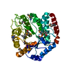
| ||||||||
|---|---|---|---|---|---|---|---|---|---|
| 1 |
| ||||||||
| Unit cell |
|
-Components
| #1: Protein | Hexosaminidase / N-acetyl-beta-glucosaminidase / Beta-N-acetylhexosaminidase Mass: 38297.523 Da / Num. of mol.: 1 Source method: isolated from a genetically manipulated source Source: (gene. exp.) Vibrio cholerae (bacteria) / Gene: Name=nagZ; OrderedLocusNames=VC0692; / Plasmid: Modified PET26B / Species (production host): Escherichia coli / Production host: Escherichia coli BL21(DE3) (bacteria) / Strain (production host): BL21(DE3) / References: UniProt: Q9KU37, beta-N-acetylhexosaminidase |
|---|---|
| #2: Water | ChemComp-HOH / Water Mass: 18.015 Da / Num. of mol.: 277 / Source method: isolated from a natural source / Formula: H2O Mass: 18.015 Da / Num. of mol.: 277 / Source method: isolated from a natural source / Formula: H2O |
-Experimental details
-Experiment
| Experiment | Method: X-RAY DIFFRACTION / Number of used crystals: 1 |
|---|
- Sample preparation
| Crystal | Density Matthews: 2.35 Å3/Da / Density % sol: 47.29 % |
|---|---|
| Crystal grow | Temperature: 293 K / Method: vapor diffusion, hanging drop / pH: 6.1 Details: 20% PEG 5000, 0.1M BisTris pH 6.5, 0.1M AmSulfate, 5% Glycerol; Cryo 30% Glycerol, 20% PEG 5000, 0.1M BisTris pH 6.5, 0.1M AmSulfate, pH 6.1, VAPOR DIFFUSION, HANGING DROP, temperature 293K |
-Data collection
| Diffraction | Mean temperature: 100 K |
|---|---|
| Diffraction source | Source: SYNCHROTRON / Site: APS  / Beamline: 31-ID / Wavelength: 0.979 Å / Beamline: 31-ID / Wavelength: 0.979 Å |
| Detector | Type: MARRESEARCH / Detector: CCD / Date: Feb 21, 2004 / Details: Vertical and Horizontal focusing mirrors |
| Radiation | Monochromator: Diamond / Protocol: SINGLE WAVELENGTH / Monochromatic (M) / Laue (L): M / Scattering type: x-ray |
| Radiation wavelength | Wavelength: 0.979 Å / Relative weight: 1 |
| Reflection | Resolution: 1.8→20 Å / Num. all: 31858 / Num. obs: 31858 / % possible obs: 100 % / Observed criterion σ(F): 0 / Observed criterion σ(I): -3 / Redundancy: 8.7 % / Biso Wilson estimate: 16.4 Å2 / Rmerge(I) obs: 0.15 / Net I/σ(I): 11.07 |
| Reflection shell | Resolution: 1.8→1.86 Å / Rmerge(I) obs: 0.283 / Mean I/σ(I) obs: 8 / Num. unique all: 3113 / % possible all: 100 |
- Processing
| Software |
| ||||||||||||||||||||||||||||||||||||||||||||||||||||||||||||||||||||||||||||||||||||||||||||||||||||||||||||||||||||||||||||||
|---|---|---|---|---|---|---|---|---|---|---|---|---|---|---|---|---|---|---|---|---|---|---|---|---|---|---|---|---|---|---|---|---|---|---|---|---|---|---|---|---|---|---|---|---|---|---|---|---|---|---|---|---|---|---|---|---|---|---|---|---|---|---|---|---|---|---|---|---|---|---|---|---|---|---|---|---|---|---|---|---|---|---|---|---|---|---|---|---|---|---|---|---|---|---|---|---|---|---|---|---|---|---|---|---|---|---|---|---|---|---|---|---|---|---|---|---|---|---|---|---|---|---|---|---|---|---|---|
| Refinement | Method to determine structure: SAD / Resolution: 1.8→20 Å / Cor.coef. Fo:Fc: 0.953 / Cor.coef. Fo:Fc free: 0.935 / SU B: 2.122 / SU ML: 0.069 / Cross valid method: THROUGHOUT / σ(F): 0 / σ(I): -3 / ESU R: 0.121 / ESU R Free: 0.114 / Stereochemistry target values: Engh & Huber / Details: HYDROGENS HAVE BEEN ADDED IN THE RIDING POSITIONS
| ||||||||||||||||||||||||||||||||||||||||||||||||||||||||||||||||||||||||||||||||||||||||||||||||||||||||||||||||||||||||||||||
| Solvent computation | Ion probe radii: 0.8 Å / Shrinkage radii: 0.8 Å / VDW probe radii: 1.2 Å / Solvent model: BABINET MODEL WITH MASK | ||||||||||||||||||||||||||||||||||||||||||||||||||||||||||||||||||||||||||||||||||||||||||||||||||||||||||||||||||||||||||||||
| Displacement parameters | Biso mean: 14.793 Å2
| ||||||||||||||||||||||||||||||||||||||||||||||||||||||||||||||||||||||||||||||||||||||||||||||||||||||||||||||||||||||||||||||
| Refinement step | Cycle: LAST / Resolution: 1.8→20 Å
| ||||||||||||||||||||||||||||||||||||||||||||||||||||||||||||||||||||||||||||||||||||||||||||||||||||||||||||||||||||||||||||||
| Refine LS restraints |
| ||||||||||||||||||||||||||||||||||||||||||||||||||||||||||||||||||||||||||||||||||||||||||||||||||||||||||||||||||||||||||||||
| LS refinement shell | Refine-ID: X-RAY DIFFRACTION / Total num. of bins used: 20
|
