 Movie
Movie Controller

[English] 日本語
 Yorodumi
- PDB-1ta0: Three-dimensional structure of a RNA-polymerase II binding protei... -
+ Open data

- Basic information
| Entry | Database: PDB / ID: 1ta0 | ||||||
|---|---|---|---|---|---|---|---|
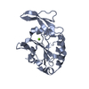
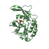
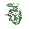
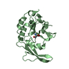
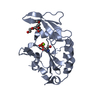
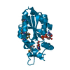
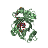
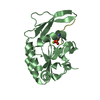
| Title | Three-dimensional structure of a RNA-polymerase II binding protein with associated ligand. | ||||||
 Components Components | Carboxy-terminal domain RNA polymerase II polypeptide A small phosphatase 1 | ||||||
 Keywords Keywords |  HYDROLASE / alpha-beta protein HYDROLASE / alpha-beta protein | ||||||
| Function / homology |  Function and homology information Function and homology informationRNA polymerase II CTD heptapeptide repeat phosphatase activity / myosin phosphatase activity / negative regulation of G1/S transition of mitotic cell cycle / protein-serine/threonine phosphatase / negative regulation of neuron differentiation / protein dephosphorylation / negative regulation of protein phosphorylation / negative regulation of neurogenesis / regulation of transcription by RNA polymerase II / extracellular exosome ...RNA polymerase II CTD heptapeptide repeat phosphatase activity / myosin phosphatase activity / negative regulation of G1/S transition of mitotic cell cycle / protein-serine/threonine phosphatase / negative regulation of neuron differentiation / protein dephosphorylation / negative regulation of protein phosphorylation / negative regulation of neurogenesis / regulation of transcription by RNA polymerase II / extracellular exosome / nucleoplasm / metal ion binding / nucleusSimilarity search - Function | ||||||
| Biological species |  Homo sapiens (human) Homo sapiens (human) | ||||||
| Method | X-RAY DIFFRACTION / SYNCHROTRON / MOLECULAR REPLACEMENT / Resolution: 2.1 Å | ||||||
 Authors Authors | Kamenski, T. / Heilmeier, S. / Meinhart, T. / Cramer, P. | ||||||
 Citation Citation | Journal: Mol.Cell / Year: 2004 Title: Structure and Mechanism of RNA Polymerase II CTD Phosphatases. Authors: Kamenski, T. / Heilmeier, S. / Meinhart, T. / Cramer, P. | ||||||
| History |
|
- Structure visualization
| Structure viewer | Molecule: MolmilJmol/JSmol |
|---|
- Downloads & links
-Download
| PDBx/mmCIF format | 1ta0.cif.gz | 50.1 KB | Display | PDBx/mmCIF format |
|---|---|---|---|---|
| PDB format | pdb1ta0.ent.gz | 38.2 KB | Display | PDB format |
| PDBx/mmJSON format | 1ta0.json.gz | Tree view | PDBx/mmJSON format | |
| Others |  Other downloads Other downloads |
-Validation report
| Arichive directory | https://data.pdbj.org/pub/pdb/validation_reports/ta/1ta0ftp://data.pdbj.org/pub/pdb/validation_reports/ta/1ta0 | HTTPS FTP |
|---|
-Related structure data


-Links
 PDBj
PDBj- Assembly
| Deposited unit | 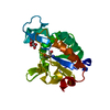
| ||||||||
|---|---|---|---|---|---|---|---|---|---|
| 1 |
| ||||||||
| Unit cell |
|
-Components
| #1: Protein | Mass: 22715.732 Da / Num. of mol.: 1 Source method: isolated from a genetically manipulated source Source: (gene. exp.) Homo sapiens (human) / Gene: CTDSP1, NIF3, NLIIF / Plasmid: pET21b / Production host:  Escherichia coli (E. coli) / Strain (production host): BL21(DE3)-RIL Escherichia coli (E. coli) / Strain (production host): BL21(DE3)-RILReferences: UniProt: Q9GZU7, protein-serine/threonine phosphatase |
|---|---|
| #2: Chemical | ChemComp-MG /   Mass: 24.305 Da / Num. of mol.: 1 / Source method: obtained synthetically / Formula: Mg Mass: 24.305 Da / Num. of mol.: 1 / Source method: obtained synthetically / Formula: Mg |
| #3: Chemical | ChemComp-CIT / Citric acid  Mass: 192.124 Da / Num. of mol.: 1 / Source method: obtained synthetically / Formula: C6H8O7 Mass: 192.124 Da / Num. of mol.: 1 / Source method: obtained synthetically / Formula: C6H8O7 |
| #4: Water | ChemComp-HOH / Water Mass: 18.015 Da / Num. of mol.: 148 / Source method: isolated from a natural source / Formula: H2O Mass: 18.015 Da / Num. of mol.: 148 / Source method: isolated from a natural source / Formula: H2O |
-Experimental details
-Experiment
| Experiment | Method: X-RAY DIFFRACTION / Number of used crystals: 1 |
|---|
- Sample preparation
| Crystal | Density Matthews: 1.3 Å3/Da / Density % sol: 54 % |
|---|---|
| Crystal grow | Temperature: 293 K / Method: vapor diffusion, hanging drop / pH: 5.6 Details: PEG 3300, ammonium acetate, citrate buffer, DTT , pH 5.6, VAPOR DIFFUSION, HANGING DROP, temperature 293K |
-Data collection
| Diffraction | Mean temperature: 100 K |
|---|---|
| Diffraction source | Source: SYNCHROTRON / Site: SLS  / Beamline: X06SA / Wavelength: 0.9919 Å / Beamline: X06SA / Wavelength: 0.9919 Å |
| Detector | Type: MARRESEARCH / Detector: CCD / Date: Feb 22, 2004 |
| Radiation | Protocol: SINGLE WAVELENGTH / Monochromatic (M) / Laue (L): M / Scattering type: x-ray |
| Radiation wavelength | Wavelength: 0.9919 Å / Relative weight: 1 |
| Reflection | Resolution: 2.1→20 Å / Num. all: 13642 / Num. obs: 13356 / % possible obs: 97.7 % / Observed criterion σ(F): 0 / Observed criterion σ(I): 0 / Redundancy: 3.8 % / Rmerge(I) obs: 0.057 |
| Reflection shell | Resolution: 2.1→2.2 Å / Rmerge(I) obs: 0.062 / Mean I/σ(I) obs: 16.8 / Num. unique all: 1520 / % possible all: 87.5 |
- Processing
| Software |
| |||||||||||||||||||||||||
|---|---|---|---|---|---|---|---|---|---|---|---|---|---|---|---|---|---|---|---|---|---|---|---|---|---|---|
| Refinement | Method to determine structure: MOLECULAR REPLACEMENT / Resolution: 2.1→20 Å / σ(F): 0 / σ(I): 0 / Stereochemistry target values: Engh & Huber
| |||||||||||||||||||||||||
| Refinement step | Cycle: LAST / Resolution: 2.1→20 Å
| |||||||||||||||||||||||||
| Refine LS restraints |
| |||||||||||||||||||||||||
| LS refinement shell | Resolution: 2.1→2.16 Å /
|
