 Movie
Movie Controller

[English] 日本語
 Yorodumi
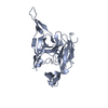
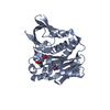
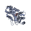
- PDB-1t7d: Crystal structure of Escherichia coli type I signal peptidase in ... -
+ Open data

- Basic information
| Entry | Database: PDB / ID: 1t7d | |||||||||
|---|---|---|---|---|---|---|---|---|---|---|
| Title | Crystal structure of Escherichia coli type I signal peptidase in complex with a lipopeptide inhibitor | |||||||||
 Components Components |
| |||||||||
 Keywords Keywords | HYDROLASE/ANTIBIOTIC /  SIGNAL PEPTIDASE / SER/LYS DYAD / HYDROLASE / LIPOPEPTIDE / ANTIBIOTIC / BIARYL BRIDGE / HYDROLASE-ANTIBIOTIC COMPLEX SIGNAL PEPTIDASE / SER/LYS DYAD / HYDROLASE / LIPOPEPTIDE / ANTIBIOTIC / BIARYL BRIDGE / HYDROLASE-ANTIBIOTIC COMPLEX | |||||||||
| Function / homology |  Function and homology informationsignal peptidase I / signal peptide processing / protein processing / peptidase activity / endopeptidase activity / serine-type endopeptidase activity / proteolysis / plasma membrane Function and homology informationsignal peptidase I / signal peptide processing / protein processing / peptidase activity / endopeptidase activity / serine-type endopeptidase activity / proteolysis / plasma membraneSimilarity search - Function | |||||||||
| Biological species |  ESCHERICHIA COLI (E. coli)STREPTOMYCES TU (bacteria) ESCHERICHIA COLI (E. coli)STREPTOMYCES TU (bacteria) | |||||||||
| Method | X-RAY DIFFRACTION / SYNCHROTRON / MOLECULAR REPLACEMENT / Resolution: 2.47 Å | |||||||||
 Authors Authors | Paetzel, M. / Goodall, J.J. / Kania, M. / Dalbey, R.E. / Page, M.G.P. | |||||||||
 Citation Citation | Journal: J.Biol.Chem. / Year: 2004 Title: Crystallographic and Biophysical Analysis of a Bacterial Signal Peptidase in Complex with a Lipopeptide Based Inhibitor. Authors: Paetzel, M. / Goodall, J.J. / Kania, M. / Dalbey, R.E. / Page, M.G.P. #1: Journal: J.Biol.Chem. / Year: 2002Title: Crystal Structure of a Bacterial Signal Peptidase Apoenzyme: Implications for Signal Peptide Binding and the Ser-Lys Dyad Mechanism Authors: Paetzel, M. / Dalbey, R.E. / Strynadka, N.C. #2: Journal: Nature / Year: 1998Title: Crystal Structure of a Bacterial Signal Peptidase in Complex with a Beta-Lactam Inhibitor. Authors: Paetzel, M. / Dalbey, R.E. / Strynadka, N.C. | |||||||||
| History |
|
- Structure visualization
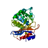
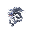
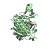
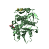
| Structure viewer | Molecule: MolmilJmol/JSmol |
|---|
- Downloads & links
-Download
| PDBx/mmCIF format | 1t7d.cif.gz | 110.7 KB | Display | PDBx/mmCIF format |
|---|---|---|---|---|
| PDB format | pdb1t7d.ent.gz | 83.1 KB | Display | PDB format |
| PDBx/mmJSON format | 1t7d.json.gz | Tree view | PDBx/mmJSON format | |
| Others |  Other downloads Other downloads |
-Validation report
| Arichive directory | https://data.pdbj.org/pub/pdb/validation_reports/t7/1t7dftp://data.pdbj.org/pub/pdb/validation_reports/t7/1t7d | HTTPS FTP |
|---|
-Related structure data
| Related structure data |  1b12S S: Starting model for refinement |
|---|---|
| Similar structure data |


-Links
 PDBj
PDBj- Assembly
| Deposited unit | 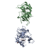
| ||||||||
|---|---|---|---|---|---|---|---|---|---|
| 1 | 
| ||||||||
| 2 | 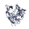
| ||||||||
| Unit cell |
| ||||||||
| Details | The biological assembly is a monomer |
-Components
| #1: Protein | / SPASE I / LEADER PEPTIDASE I Mass: 28079.814 Da / Num. of mol.: 2 / Fragment: RESIDUES 76-324 Source method: isolated from a genetically manipulated source Source: (gene. exp.) ESCHERICHIA COLI (E. coli) / Gene: LEPB / Plasmid: PET3D / Production host: ESCHERICHIA COLI BL21(DE3) (bacteria) / Strain (production host): BL21(DE3) / References: UniProt: P00803, signal peptidase I#2: Protein/peptide | /   Type: Lipopeptide / Class: Antibiotic / Mass: 644.674 Da / Num. of mol.: 2 / Source method: isolated from a natural source Type: Lipopeptide / Class: Antibiotic / Mass: 644.674 Da / Num. of mol.: 2 / Source method: isolated from a natural sourceDetails: ARYLOMYCIN A2 IS A BIARYL-BRIDGED LIPOPEPTIDE. THE SCAFFOLD IS MADE OF TWO PARTS: (1) N-TERM EXOCYCLIC TRIPEPTIDE (2) A TRICYCLIC PEPTIDE, WITH [3,3] BIARYL BOND BETWEEN RESIDUE 4 AND 6 AN ...Details: ARYLOMYCIN A2 IS A BIARYL-BRIDGED LIPOPEPTIDE. THE SCAFFOLD IS MADE OF TWO PARTS: (1) N-TERM EXOCYCLIC TRIPEPTIDE (2) A TRICYCLIC PEPTIDE, WITH [3,3] BIARYL BOND BETWEEN RESIDUE 4 AND 6 AN ISO-C12 FATTY ACID IS LINKED TO RESIDUE 1. Source: (natural) STREPTOMYCES TU (bacteria) / References: NOR: NOR01115, ARYLOMYCIN A2#3: Chemical | ChemComp-M12 /   Type: Lipopeptide / Class: Antibiotic / Mass: 200.318 Da / Num. of mol.: 2 / Source method: obtained synthetically / Formula: C12H24O2 Type: Lipopeptide / Class: Antibiotic / Mass: 200.318 Da / Num. of mol.: 2 / Source method: obtained synthetically / Formula: C12H24O2Details: ARYLOMYCIN A2 IS A BIARYL-BRIDGED LIPOPEPTIDE. THE SCAFFOLD IS MADE OF TWO PARTS: (1) N-TERM EXOCYCLIC TRIPEPTIDE (2) A TRICYCLIC PEPTIDE, WITH [3,3] BIARYL BOND BETWEEN RESIDUE 4 AND 6 AN ...Details: ARYLOMYCIN A2 IS A BIARYL-BRIDGED LIPOPEPTIDE. THE SCAFFOLD IS MADE OF TWO PARTS: (1) N-TERM EXOCYCLIC TRIPEPTIDE (2) A TRICYCLIC PEPTIDE, WITH [3,3] BIARYL BOND BETWEEN RESIDUE 4 AND 6 AN ISO-C12 FATTY ACID IS LINKED TO RESIDUE 1. References: ARYLOMYCIN A2#4: Water | ChemComp-HOH / | Water Mass: 18.015 Da / Num. of mol.: 265 / Source method: isolated from a natural source / Formula: H2O Mass: 18.015 Da / Num. of mol.: 265 / Source method: isolated from a natural source / Formula: H2OCompound details | ARYLOMYCIN A2 IS A CYCLIC LIPOHEXAPEPTIDE, A MEMBER OF THE ARYLOMYCIN FAMILY. ALL MEMBERS HAVE A ...ARYLOMYCIN | Sequence details | THE PROTEIN IS A SOLUBLE CATALYTICALLY ACTIVE FRAGMENT OF THE ESCHERICHIA COLI TYPE I SIGNAL ...THE PROTEIN IS A SOLUBLE CATALYTICA | |
|---|
-Experimental details
-Experiment
| Experiment | Method: X-RAY DIFFRACTION / Number of used crystals: 1 |
|---|
- Sample preparation
| Crystal | Density Matthews: 2.8 Å3/Da / Density % sol: 56 % |
|---|---|
| Crystal grow | pH: 6 Details: 0.5% TRITON X-100, 15% PEG 4000, 20% PROPANOL, 0.1 M SODIUM CITRATE, PH 6.0, VAPOR DIFFUSION, SITTING DROP, TEMPERATURE 298K |
-Data collection
| Diffraction | Mean temperature: 100 K |
|---|---|
| Diffraction source | Source: SYNCHROTRON / Site: NSLS  / Beamline: X8C / Wavelength: 1 / Beamline: X8C / Wavelength: 1 |
| Detector | Type: ADSC QUANTUM 4 / Detector: CCD / Date: May 5, 2003 |
| Radiation | Monochromator: GRAPHITE / Protocol: SINGLE WAVELENGTH / Monochromatic (M) / Laue (L): M / Scattering type: x-ray |
| Radiation wavelength | Wavelength: 1 Å / Relative weight: 1 |
| Reflection | Resolution: 2.47→40 Å / Num. obs: 21589 / % possible obs: 90.9 % / Biso Wilson estimate: 45.3 Å2 / Rmerge(I) obs: 0.077 / Net I/σ(I): 19.4 |
| Reflection shell | Resolution: 2.47→2.56 Å / Rmerge(I) obs: 0.183 / Mean I/σ(I) obs: 8 / % possible all: 86.4 |
- Processing
| Software |
| ||||||||||||||||||||||||||||||||||||||||||||||||||||||||||||||||||||||||||||||||
|---|---|---|---|---|---|---|---|---|---|---|---|---|---|---|---|---|---|---|---|---|---|---|---|---|---|---|---|---|---|---|---|---|---|---|---|---|---|---|---|---|---|---|---|---|---|---|---|---|---|---|---|---|---|---|---|---|---|---|---|---|---|---|---|---|---|---|---|---|---|---|---|---|---|---|---|---|---|---|---|---|---|
| Refinement | Method to determine structure: MOLECULAR REPLACEMENT Starting model: PDB ENTRY 1B12 Resolution: 2.47→29.54 Å / Rfactor Rfree error: 0.006 / Data cutoff high absF: 2098721.28 / Data cutoff low absF: 0 / Isotropic thermal model: RESTRAINED / Cross valid method: THROUGHOUT / σ(F): 0 / Stereochemistry target values: ENGH & HUBER
| ||||||||||||||||||||||||||||||||||||||||||||||||||||||||||||||||||||||||||||||||
| Solvent computation | Solvent model: FLAT MODEL / Bsol: 49.11 Å2 / ksol: 0.33 e/Å3 | ||||||||||||||||||||||||||||||||||||||||||||||||||||||||||||||||||||||||||||||||
| Displacement parameters | Biso mean: 49.5 Å2
| ||||||||||||||||||||||||||||||||||||||||||||||||||||||||||||||||||||||||||||||||
| Refine analyze |
| ||||||||||||||||||||||||||||||||||||||||||||||||||||||||||||||||||||||||||||||||
| Refinement step | Cycle: LAST / Resolution: 2.47→29.54 Å
| ||||||||||||||||||||||||||||||||||||||||||||||||||||||||||||||||||||||||||||||||
| Refine LS restraints |
| ||||||||||||||||||||||||||||||||||||||||||||||||||||||||||||||||||||||||||||||||
| LS refinement shell | Resolution: 2.47→2.62 Å / Rfactor Rfree error: 0.02 / Total num. of bins used: 6
| ||||||||||||||||||||||||||||||||||||||||||||||||||||||||||||||||||||||||||||||||
| Xplor file |
|
