[English] 日本語
Yorodumi
- PDB-1sq0: Crystal Structure of the Complex of the Wild-type Von Willebrand ... -

+
Open data


ID or keywords:

Loading...

-
Basic information

Entry
Database: PDB / ID: 1sq0
TitleCrystal Structure of the Complex of the Wild-type Von Willebrand Factor A1 domain and Glycoprotein Ib alpha at 2.6 Angstrom Resolution
Components
  • Platelet glycoprotein Ib alpha chain (Glycoprotein Ibalpha) (GP-Ib alpha) (GPIbA) (GPIb-alpha) (CD42B-alpha) (CD42B) [Contains: Glycocalicin]
  • Von Willebrand factor (vWF) [Contains: Von Willebrand antigen II]
Keywords
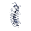
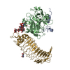
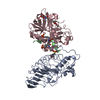
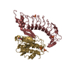
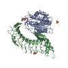
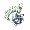
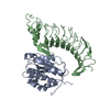
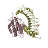
BLOOD CLOTTING / Leucine Rich Repeat (LRR) / right-handed beta-alpha superhelix) / Integrin A (or I) domain fold
Function / homology
Function and homology information


thrombin-activated receptor activity / glycoprotein Ib-IX-V complex / Weibel-Palade body / Defective F8 binding to von Willebrand factor / positive regulation of leukocyte tethering or rolling / blood coagulation, intrinsic pathway / hemostasis / Defective F9 activation / Platelet Adhesion to exposed collagen / platelet alpha granule ...thrombin-activated receptor activity / glycoprotein Ib-IX-V complex / Weibel-Palade body / Defective F8 binding to von Willebrand factor / positive regulation of leukocyte tethering or rolling / blood coagulation, intrinsic pathway / hemostasis / Defective F9 activation / Platelet Adhesion to exposed collagen / platelet alpha granule / positive regulation of platelet activation / positive regulation of intracellular signal transduction / megakaryocyte development / GP1b-IX-V activation signalling / p130Cas linkage to MAPK signaling for integrins / cell-substrate adhesion / regulation of blood coagulation / immunoglobulin binding / Defective F8 cleavage by thrombin / Platelet Aggregation (Plug Formation) / GRB2:SOS provides linkage to MAPK signaling for Integrins / Integrin cell surface interactions / release of sequestered calcium ion into cytosol / collagen binding / fibrinolysis / Intrinsic Pathway of Fibrin Clot Formation / extracellular matrix / Integrin signaling / platelet alpha granule lumen / Signaling by high-kinase activity BRAF mutants / RUNX1 regulates genes involved in megakaryocyte differentiation and platelet function / MAP2K and MAPK activation / cell morphogenesis / platelet activation / response to wounding / Signaling by RAF1 mutants / Signaling by moderate kinase activity BRAF mutants / Paradoxical activation of RAF signaling by kinase inactive BRAF / Signaling downstream of RAS mutants / Signaling by BRAF and RAF1 fusions / blood coagulation / integrin binding / Platelet degranulation / protein-folding chaperone binding / collagen-containing extracellular matrix / protease binding / cell surface receptor signaling pathway / cell adhesion / external side of plasma membrane / cell surface / endoplasmic reticulum / extracellular space / extracellular exosome / extracellular region / membrane / identical protein binding / plasma membrane
Similarity search - Function
von Willebrand factor, VWA N-terminal domain / Von Willebrand factor / VWA N-terminal / C8 domain / Uncharacterised domain, cysteine-rich / C8 / von Willebrand factor, type D domain / von Willebrand factor type D domain / VWFD domain profile. / von Willebrand factor (vWF) type D domain ...von Willebrand factor, VWA N-terminal domain / Von Willebrand factor / VWA N-terminal / C8 domain / Uncharacterised domain, cysteine-rich / C8 / von Willebrand factor, type D domain / von Willebrand factor type D domain / VWFD domain profile. / von Willebrand factor (vWF) type D domain / C-terminal cystine knot signature. / von Willebrand factor (vWF) type C domain / Trypsin Inhibitor-like, cysteine rich domain / Serine protease inhibitor-like superfamily / Trypsin Inhibitor like cysteine rich domain / C-terminal cystine knot domain profile. / Leucine rich repeat N-terminal domain / Cystine knot, C-terminal / C-terminal cystine knot-like domain (CTCK) / von Willebrand factor type C domain / Leucine-rich repeat N-terminal domain / Leucine rich repeat N-terminal domain / VWFC domain signature. / VWFC domain profile. / von Willebrand factor (vWF) type C domain / VWFC domain / Leucine-rich repeats, bacterial type / von Willebrand factor, type A domain / Cysteine-rich flanking region, C-terminal / Leucine rich repeat C-terminal domain / Leucine-rich repeat, LRR (right-handed beta-alpha superhelix) / Ribonuclease Inhibitor / von Willebrand factor type A domain / Alpha-Beta Horseshoe / von Willebrand factor (vWF) type A domain / VWFA domain profile. / Leucine rich repeat / Leucine-rich repeat, typical subtype / Leucine-rich repeats, typical (most populated) subfamily / von Willebrand factor, type A / Leucine-rich repeat profile. / von Willebrand factor A-like domain superfamily / Leucine-rich repeat / Leucine-rich repeat domain superfamily / Rossmann fold / 3-Layer(aba) Sandwich / Alpha Beta
Similarity search - Domain/homology
von Willebrand factor / Platelet glycoprotein Ib alpha chain
Similarity search - Component
Biological speciesHomo sapiens (human)
MethodX-RAY DIFFRACTION / SYNCHROTRON / MAD / Resolution: 2.6 Å
AuthorsDumas, J.J. / Kumar, R. / McDonagh, T. / Sullivan, F. / Stahl, M.L. / Somers, W.S. / Mosyak, L.
Citation
Journal: J.Biol.Chem. / Year: 2004
Title: Crystal structure of the wild-type von Willebrand factor A1-glycoprotein Ibalpha complex reveals conformation differences with a complex bearing von Willebrand disease mutations
Authors: Dumas, J.J. / Kumar, R. / McDonagh, T. / Sullivan, F. / Stahl, M.L. / Somers, W.S. / Mosyak, L.
#1: Journal: Science / Year: 2003
Title: Crystal Structure of the GpIb(alpha)-Thrombin Complex Essential for Platelet Aggregation
Authors: Dumas, J.J. / Kumar, R. / Seehra, J. / Somers, W.S. / Mosyak, L.
History
DepositionMar 17, 2004Deposition site: RCSB / Processing site: RCSB
Revision 1.0Apr 13, 2004Provider: repository / Type: Initial release
Revision 1.1Apr 29, 2008Group: Version format compliance
Revision 1.2Jul 13, 2011Group: Version format compliance
Revision 1.3Oct 27, 2021Group: Database references / Category: database_2 / struct_ref_seq_dif
Item: _database_2.pdbx_DOI / _database_2.pdbx_database_accession / _struct_ref_seq_dif.details
Remark 999SEQUENCE ASN RESIDUES AT POSITIONS 21 AND 159 OF CHAIN B, WHICH ARE SITES FOR N-LINKED ...SEQUENCE ASN RESIDUES AT POSITIONS 21 AND 159 OF CHAIN B, WHICH ARE SITES FOR N-LINKED GLYCOSYLATION, WERE MUTATED TO ASP.

-
Structure visualization

Structure viewerMolecule:
MolmilJmol/JSmol

Downloads & links

-
Assembly

Deposited unit
A: Von Willebrand factor (vWF) [Contains: Von Willebrand antigen II]
B: Platelet glycoprotein Ib alpha chain (Glycoprotein Ibalpha) (GP-Ib alpha) (GPIbA) (GPIb-alpha) (CD42B-alpha) (CD42B) [Contains: Glycocalicin]


Theoretical massNumber of molelcules
Total (without water)56,5512
Polymers56,5512
Non-polymers00
Water5,729318
1


  • Idetical with deposited unit
  • defined by author
TypeNameSymmetry operationNumber
identity operation1_555x,y,z1
Unit cell
Length a, b, c (Å)72.497, 116.104, 67.962
Angle α, β, γ (deg.)90.00, 90.00, 90.00
Int Tables number18
Space group name H-MP21212

-
Components

#1: Protein Von Willebrand factor (vWF) [Contains: Von Willebrand antigen II]


Mass: 24466.361 Da / Num. of mol.: 1 / Fragment: A1
Source method: isolated from a genetically manipulated source
Source: (gene. exp.) Homo sapiens (human) / Gene: VWF, F8VWF / Species (production host): Escherichia coli / Production host: Escherichia coli BL21(DE3) (bacteria) / Strain (production host): BL21DE3 / References: UniProt: P04275
#2: Protein Platelet glycoprotein Ib alpha chain (Glycoprotein Ibalpha) (GP-Ib alpha) (GPIbA) (GPIb-alpha) (CD42B-alpha) (CD42B) [Contains: Glycocalicin]


Mass: 32084.498 Da / Num. of mol.: 1 / Fragment: Glycoprotein Ib-(alpha) / Mutation: N21D, N159D
Source method: isolated from a genetically manipulated source
Source: (gene. exp.) Homo sapiens (human) / Gene: GP1BA / Cell (production host): CHO / Production host: Cricetulus griseus (Chinese hamster) / References: UniProt: P07359
#3: Water ChemComp-HOH / water / Water


Mass: 18.015 Da / Num. of mol.: 318 / Source method: isolated from a natural source / Formula: H2O

-
Experimental details

-
Experiment

ExperimentMethod: X-RAY DIFFRACTION / Number of used crystals: 2

-
Sample preparation

CrystalDensity Matthews: 2.85 Å3/Da / Density % sol: 56.5 %
Crystal growTemperature: 291 K / Method: vapor diffusion, hanging drop / pH: 8
Details: PEG-3350; potassium thiocyanate, pH 8.0, VAPOR DIFFUSION, HANGING DROP, temperature 291K

-
Data collection

Diffraction
IDMean temperature (K)Crystal-ID
11001
21
Diffraction source
SourceSiteBeamlineIDWavelength (Å)
SYNCHROTRONALS 5.0.210.97942, 0.97959, 0.9686
SYNCHROTRONALS 5.0.221
Detector
TypeIDDetectorDate
ADSC QUANTUM 41CCDAug 31, 2002
ADSC QUANTUM 42CCDMay 18, 2002
Radiation
IDMonochromatorProtocolMonochromatic (M) / Laue (L)Scattering typeWavelength-ID
1Double-crystal, Si(111)MADMx-ray1
2Mx-ray1
Radiation wavelength
IDWavelength (Å)Relative weight
10.979421
20.979591
30.96861
411
ReflectionResolution: 2.6→49.58 Å / Num. obs: 17150 / Observed criterion σ(I): 0 / Biso Wilson estimate: 28.5 Å2
Reflection shellResolution: 2.6→2.75 Å / % possible all: 84

-
Processing

Software
NameVersionClassification
CNS1.1refinement
DENZOdata reduction
CCP4(SCALA)data scaling
SHELXSphasing
RefinementMethod to determine structure: MAD / Resolution: 2.6→49.58 Å / Rfactor Rfree error: 0.006 / Data cutoff high absF: 132767.56 / Data cutoff low absF: 0 / Isotropic thermal model: RESTRAINED / Cross valid method: THROUGHOUT / σ(F): 0
Details: DISORDERED RESIDUES IN CHAIN A INCLUDE N-TERMINAL RESIDUES 496-5 AND C-TERMINAL RESIDUES 704-709. IN CHAIN A, SIDE CHAIN FOR LYS IS DISORDERED. DISORDERED RESIDUES IN CHAIN B INCLUDE C-TERMINAL RESIDUES 266-288.
RfactorNum. reflection% reflectionSelection details
Rfree0.239 1685 9.8 %RANDOM
Rwork0.19 ---
obs0.19 17150 93.9 %-
all-17150 --
Solvent computationSolvent model: FLAT MODEL / Bsol: 34.4256 Å2 / ksol: 0.305156 e/Å3
Displacement parametersBiso mean: 40.6 Å2
Baniso -1Baniso -2Baniso -3
1--2.42 Å20 Å20 Å2
2---10.55 Å20 Å2
3---12.98 Å2
Refine analyze
FreeObs
Luzzati coordinate error0.37 Å0.28 Å
Luzzati d res low-5 Å
Luzzati sigma a0.42 Å0.36 Å
Refinement stepCycle: LAST / Resolution: 2.6→49.58 Å
ProteinNucleic acidLigandSolventTotal
Num. atoms3664 0 0 318 3982
Refine LS restraints
Refine-IDTypeDev idealDev ideal target
X-RAY DIFFRACTIONc_bond_d0.006
X-RAY DIFFRACTIONc_bond_d_na
X-RAY DIFFRACTIONc_bond_d_prot
X-RAY DIFFRACTIONc_angle_d
X-RAY DIFFRACTIONc_angle_d_na
X-RAY DIFFRACTIONc_angle_d_prot
X-RAY DIFFRACTIONc_angle_deg1.3
X-RAY DIFFRACTIONc_angle_deg_na
X-RAY DIFFRACTIONc_angle_deg_prot
X-RAY DIFFRACTIONc_dihedral_angle_d26
X-RAY DIFFRACTIONc_dihedral_angle_d_na
X-RAY DIFFRACTIONc_dihedral_angle_d_prot
X-RAY DIFFRACTIONc_improper_angle_d0.87
X-RAY DIFFRACTIONc_improper_angle_d_na
X-RAY DIFFRACTIONc_improper_angle_d_prot
X-RAY DIFFRACTIONc_mcbond_it1.561.5
X-RAY DIFFRACTIONc_mcangle_it2.622
X-RAY DIFFRACTIONc_scbond_it2.342
X-RAY DIFFRACTIONc_scangle_it3.582.5
LS refinement shellResolution: 2.6→2.76 Å / Rfactor Rfree error: 0.02 / Total num. of bins used: 6
RfactorNum. reflection% reflection
Rfree0.322 248 9.9 %
Rwork0.281 2261 -
obs--84 %
Xplor file
Refine-IDSerial noParam fileTopol file
X-RAY DIFFRACTION1PROTEIN_REP.PARAMPROTEIN.TOP
X-RAY DIFFRACTION2WATER_REP.PARAMWATER.TOP
X-RAY DIFFRACTION4ION.PARAMION.TOP

+
About Yorodumi

-
News

-
Feb 9, 2022. New format data for meta-information of EMDB entries

New format data for meta-information of EMDB entries

  • Version 3 of the EMDB header file is now the official format.
  • The previous official version 1.9 will be removed from the archive.

Related info.:EMDB header

External links:wwPDB to switch to version 3 of the EMDB data model

-
Aug 12, 2020. Covid-19 info

Covid-19 info

URL: https://pdbj.org/emnavi/covid19.php

New page: Covid-19 featured information page in EM Navigator.

Related info.:Covid-19 info / Mar 5, 2020. Novel coronavirus structure data

+
Mar 5, 2020. Novel coronavirus structure data

Novel coronavirus structure data

Related info.:Yorodumi Speices / Aug 12, 2020. Covid-19 info

External links:COVID-19 featured content - PDBj / Molecule of the Month (242):Coronavirus Proteases

+
Jan 31, 2019. EMDB accession codes are about to change! (news from PDBe EMDB page)

EMDB accession codes are about to change! (news from PDBe EMDB page)

  • The allocation of 4 digits for EMDB accession codes will soon come to an end. Whilst these codes will remain in use, new EMDB accession codes will include an additional digit and will expand incrementally as the available range of codes is exhausted. The current 4-digit format prefixed with “EMD-” (i.e. EMD-XXXX) will advance to a 5-digit format (i.e. EMD-XXXXX), and so on. It is currently estimated that the 4-digit codes will be depleted around Spring 2019, at which point the 5-digit format will come into force.
  • The EM Navigator/Yorodumi systems omit the EMD- prefix.

Related info.:Q: What is EMD? / ID/Accession-code notation in Yorodumi/EM Navigator

External links:EMDB Accession Codes are Changing Soon! / Contact to PDBj

+
Jul 12, 2017. Major update of PDB

Major update of PDB

  • wwPDB released updated PDB data conforming to the new PDBx/mmCIF dictionary.
  • This is a major update changing the version number from 4 to 5, and with Remediation, in which all the entries are updated.
  • In this update, many items about electron microscopy experimental information are reorganized (e.g. em_software).
  • Now, EM Navigator and Yorodumi are based on the updated data.

External links:wwPDB Remediation / Enriched Model Files Conforming to OneDep Data Standards Now Available in the PDB FTP Archive

-
Yorodumi

Thousand views of thousand structures

  • Yorodumi is a browser for structure data from EMDB, PDB, SASBDB, etc.
  • This page is also the successor to EM Navigator detail page, and also detail information page/front-end page for Omokage search.
  • The word "yorodu" (or yorozu) is an old Japanese word meaning "ten thousand". "mi" (miru) is to see.

Related info.:EMDB / PDB / SASBDB / Comparison of 3 databanks / Yorodumi Search / Aug 31, 2016. New EM Navigator & Yorodumi / Yorodumi Papers / Jmol/JSmol / Function and homology information / Changes in new EM Navigator and Yorodumi

Read more