 Movie
Movie Controller

[English] 日本語
 Yorodumi
- PDB-1spv: Crystal Structure of the Putative Phosphatase of Escherichia coli... -
+ Open data

- Basic information
| Entry | Database: PDB / ID: 1spv | ||||||
|---|---|---|---|---|---|---|---|
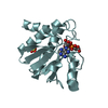
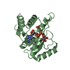
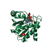
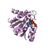
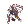
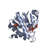
| Title | Crystal Structure of the Putative Phosphatase of Escherichia coli, Northeast Structural Genomoics Target ER58 | ||||||
 Components Components | putative polyprotein/phosphatase | ||||||
 Keywords Keywords |  STRUCTURAL GENOMICS / UNKNOWN FUNCTION / Structural Genomoics / alpha/beta monomeric protein / PSI / Protein Structure Initiative / Northeast Structural Genomics Consortium / NESG STRUCTURAL GENOMICS / UNKNOWN FUNCTION / Structural Genomoics / alpha/beta monomeric protein / PSI / Protein Structure Initiative / Northeast Structural Genomics Consortium / NESG | ||||||
| Function / homology |  Function and homology information Function and homology informationO-acetyl-ADP-ribose deacetylase / regulation of single-species biofilm formation on inanimate substrate / O-acetyl-ADP-ribose deacetylase activity / purine nucleoside binding / purine nucleoside metabolic process / endoribonuclease inhibitor activity / response to antibiotic / enzyme bindingSimilarity search - Function | ||||||
| Biological species |  Escherichia coli (E. coli) Escherichia coli (E. coli) | ||||||
| Method | X-RAY DIFFRACTION / SYNCHROTRON / SAD / Resolution: 2 Å | ||||||
 Authors Authors | Forouhar, F. / Lee, I. / Vorobiev, S.M. / Xiao, R. / Acton, T.B. / Montelione, G.T. / Hunt, J.F. / Tong, L. / Northeast Structural Genomics Consortium (NESG) | ||||||
 Citation Citation | Journal: To be Published Title: Crystal Structure of the Putative Phosphatase of Escherichia coli, Northeast Structural Genomoics Target ER58 Authors: Forouhar, F. / Lee, I. / Vorobiev, S.M. / Xiao, R. / Acton, T.B. / Montelione, G.T. / Hunt, J.F. / Tong, L. | ||||||
| History |
|
- Structure visualization
| Structure viewer | Molecule: MolmilJmol/JSmol |
|---|
- Downloads & links
-Download
| PDBx/mmCIF format | 1spv.cif.gz | 42.9 KB | Display | PDBx/mmCIF format |
|---|---|---|---|---|
| PDB format | pdb1spv.ent.gz | 33.2 KB | Display | PDB format |
| PDBx/mmJSON format | 1spv.json.gz | Tree view | PDBx/mmJSON format | |
| Others |  Other downloads Other downloads |
-Validation report
| Arichive directory | https://data.pdbj.org/pub/pdb/validation_reports/sp/1spvftp://data.pdbj.org/pub/pdb/validation_reports/sp/1spv | HTTPS FTP |
|---|
-Related structure data
| Similar structure data | |
|---|---|
| Other databases |
-Links
 PDBj
PDBj- Assembly
| Deposited unit | 
| ||||||||
|---|---|---|---|---|---|---|---|---|---|
| 1 |
| ||||||||
| Unit cell |
|
-Components
| #1: Protein | Mass: 19904.217 Da / Num. of mol.: 1 Source method: isolated from a genetically manipulated source Source: (gene. exp.) Escherichia coli (E. coli) / Plasmid: pET15 / Production host: Escherichia coli (E. coli) / Strain (production host): BL21(DE3)+Magic / References: UniProt: P0A8D6 |
|---|---|
| #2: Chemical | ChemComp-MES / MES (buffer)  Mass: 195.237 Da / Num. of mol.: 1 / Source method: obtained synthetically / Formula: C6H13NO4S / Comment: pH buffer*YM Mass: 195.237 Da / Num. of mol.: 1 / Source method: obtained synthetically / Formula: C6H13NO4S / Comment: pH buffer*YM |
| #3: Water | ChemComp-HOH / Water Mass: 18.015 Da / Num. of mol.: 98 / Source method: isolated from a natural source / Formula: H2O Mass: 18.015 Da / Num. of mol.: 98 / Source method: isolated from a natural source / Formula: H2O |
-Experimental details
-Experiment
| Experiment | Method: X-RAY DIFFRACTION / Number of used crystals: 1 |
|---|
- Sample preparation
| Crystal | Density Matthews: 2.26 Å3/Da / Density % sol: 45.13 % |
|---|---|
| Crystal grow | Temperature: 277 K / Method: vapor diffusion, hanging drop Details: Well solution: 12% PEG 20K, 100mM MES pH 5.75, 30mM KH2PO4. Protein Solution: 10mM Tris pH 7.5, 5mM DTT, 100mM NaCl, VAPOR DIFFUSION, HANGING DROP, temperature 277K |
-Data collection
| Diffraction | Mean temperature: 100 K |
|---|---|
| Diffraction source | Source: SYNCHROTRON / Site: NSLS  / Beamline: X4A / Wavelength: 0.97927 Å / Beamline: X4A / Wavelength: 0.97927 Å |
| Detector | Type: ADSC QUANTUM 4 / Detector: CCD / Date: Mar 8, 2004 / Details: mirrors |
| Radiation | Protocol: SINGLE WAVELENGTH / Monochromatic (M) / Laue (L): M / Scattering type: x-ray |
| Radiation wavelength | Wavelength: 0.97927 Å / Relative weight: 1 |
| Reflection | Resolution: 2→29.29 Å / Num. obs: 22088 / % possible obs: 99.2 % / Observed criterion σ(F): 2 / Observed criterion σ(I): 2 / Redundancy: 4.15 % / Biso Wilson estimate: 9.5 Å2 / Rmerge(I) obs: 0.06 / Rsym value: 0.065 / Net I/σ(I): 18.84 |
| Reflection shell | Resolution: 2→2.07 Å / Redundancy: 3.2 % / Rmerge(I) obs: 0.11 / Mean I/σ(I) obs: 7.7 / Num. unique all: 2214 / Rsym value: 0.116 / % possible all: 97.3 |
- Processing
| Software |
| |||||||||||||||||||||||||
|---|---|---|---|---|---|---|---|---|---|---|---|---|---|---|---|---|---|---|---|---|---|---|---|---|---|---|
| Refinement | Method to determine structure: SAD / Resolution: 2→29.29 Å / Isotropic thermal model: anisotropic / Cross valid method: THROUGHOUT / σ(F): 2 / σ(I): 2 / Stereochemistry target values: Engh & Huber / Details: Used weighted full matrix least squares procedure
| |||||||||||||||||||||||||
| Displacement parameters | Biso mean: 22.1 Å2
| |||||||||||||||||||||||||
| Refine analyze |
| |||||||||||||||||||||||||
| Refinement step | Cycle: LAST / Resolution: 2→29.29 Å
| |||||||||||||||||||||||||
| Refine LS restraints |
| |||||||||||||||||||||||||
| LS refinement shell | Resolution: 2→2.13 Å / Rfactor Rfree error: 0.018
|
