 Movie
Movie Controller

[English] 日本語
 Yorodumi
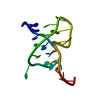
- PDB-1spk: Solution Structure of RSGI RUH-010, an SH3 Domain from Mouse cDNA -
+ Open data

- Basic information
| Entry | Database: PDB / ID: 1spk | ||||||
|---|---|---|---|---|---|---|---|
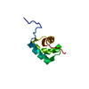
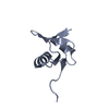
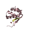
| Title | Solution Structure of RSGI RUH-010, an SH3 Domain from Mouse cDNA | ||||||
 Components Components | RIKEN cDNA 1300006M19 | ||||||
 Keywords Keywords |  STRUCTURAL GENOMICS / UNKNOWN FUNCTION / SH3 domain / five-stranded barrel / mouse cDNA / RIKEN Structural Genomics/Proteomics Initiative / RSGI STRUCTURAL GENOMICS / UNKNOWN FUNCTION / SH3 domain / five-stranded barrel / mouse cDNA / RIKEN Structural Genomics/Proteomics Initiative / RSGI | ||||||
| Function / homology |  Function and homology information Function and homology information: / regulation of insulin receptor signaling pathway / plasma membrane organization / actin crosslink formation / proline-rich region binding / positive regulation of actin filament polymerization / actin filament bundle assembly / response to bacterium / actin cytoskeleton / actin binding ...: / regulation of insulin receptor signaling pathway / plasma membrane organization / actin crosslink formation / proline-rich region binding / positive regulation of actin filament polymerization / actin filament bundle assembly / response to bacterium / actin cytoskeleton / actin binding / nucleoplasm / plasma membrane / cytosolSimilarity search - Function | ||||||
| Biological species |  Mus musculus (house mouse) Mus musculus (house mouse) | ||||||
| Method | SOLUTION NMR / simulated annealing, torsion angle dynamics | ||||||
 Authors Authors | Suzuki, Y. / Abe, T. / Hirota, H. / Hayashi, F. / Yokoyama, S. / RIKEN Structural Genomics/Proteomics Initiative (RSGI) | ||||||
 Citation Citation | Journal: To be Published Title: Solution Structure of RSGI RUH-010, an SH3 Domain from Mouse cDNA Authors: Suzuki, Y. / Abe, T. / Hirota, H. / Hayashi, F. / Yokoyama, S. | ||||||
| History |
| ||||||
| Remark 650 | HELIX DETERMINATION METHOD: AUTHOR DETERMINED | ||||||
| Remark 700 | SHEET DETERMINATION METHOD: AUTHOR DETERMINED |
- Structure visualization
| Structure viewer | Molecule: MolmilJmol/JSmol |
|---|
- Downloads & links
-Download
| PDBx/mmCIF format | 1spk.cif.gz | 412.2 KB | Display | PDBx/mmCIF format |
|---|---|---|---|---|
| PDB format | pdb1spk.ent.gz | 358.7 KB | Display | PDB format |
| PDBx/mmJSON format | 1spk.json.gz | Tree view | PDBx/mmJSON format | |
| Others |  Other downloads Other downloads |
-Validation report
| Arichive directory | https://data.pdbj.org/pub/pdb/validation_reports/sp/1spkftp://data.pdbj.org/pub/pdb/validation_reports/sp/1spk | HTTPS FTP |
|---|
-Related structure data
| Similar structure data | |
|---|---|
| Other databases |


-Links
 PDBj
PDBj
- Assembly
| Deposited unit | 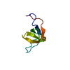
| |||||||||
|---|---|---|---|---|---|---|---|---|---|---|
| 1 |
| |||||||||
| NMR ensembles |
|
-Components
| #1: Protein | Mass: 7664.490 Da / Num. of mol.: 1 Fragment: Putative SH3 domain(Residues 343-401 in seq. db No.) Source method: isolated from a genetically manipulated source Source: (gene. exp.) Mus musculus (house mouse) / Description: Cell-free protein synthesis / Gene: RIKEN cDNA 1300006M19 / Organ: liver / Plasmid: P030120-88 / References: UniProt: Q9DBJ3 |
|---|
-Experimental details
-Experiment
| Experiment | Method: SOLUTION NMR | ||||||||||||
|---|---|---|---|---|---|---|---|---|---|---|---|---|---|
| NMR experiment |
| ||||||||||||
| NMR details | Text: The structure was determined using triple-resonance NMR spectroscopy. |
- Sample preparation
| Details | Contents: 1.0mM protein; 20mM sodium phosphate; 100mM NaCl; 1mM d-DTT; 0.02% NaN3; 10% D2O Solvent system: 90% H2O/10% D2O |
|---|---|
| Sample conditions | Ionic strength: 100mM / pH: 6.0 / Pressure: ambient / Temperature: 298 K |
-NMR measurement
| Radiation | Protocol: SINGLE WAVELENGTH / Monochromatic (M) / Laue (L): M |
|---|---|
| Radiation wavelength | Relative weight: 1 |
| NMR spectrometer | Type: Varian INOVA / Manufacturer: Varian / Model: INOVA / Field strength: 800 MHz |
- Processing
| NMR software |
| ||||||||||||||||||||||||
|---|---|---|---|---|---|---|---|---|---|---|---|---|---|---|---|---|---|---|---|---|---|---|---|---|---|
| Refinement | Method: simulated annealing, torsion angle dynamics / Software ordinal: 1 | ||||||||||||||||||||||||
| NMR representative | Selection criteria: lowest energy, target function | ||||||||||||||||||||||||
| NMR ensemble | Conformer selection criteria: structures with the least restraint violations, structures with the lowest energy, target function Conformers calculated total number: 100 / Conformers submitted total number: 20 |
