 Movie
Movie Controller

[English] 日本語
 Yorodumi
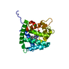
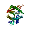
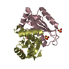
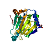
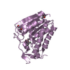
- PDB-1sl7: Crystal structure of calcium-loaded apo-obelin from Obelia longissima -
+ Open data

- Basic information
| Entry | Database: PDB / ID: 1sl7 | ||||||
|---|---|---|---|---|---|---|---|
| Title | Crystal structure of calcium-loaded apo-obelin from Obelia longissima | ||||||
 Components Components | Obelin | ||||||
 Keywords Keywords |  LUMINESCENT PROTEIN / Photoprotein / obelin / bioluminescence / calcium binding / EF-hand / aequorin / fluorescence / Structural Genomics / PSI / Protein Structure Initiative / Southeast Collaboratory for Structural Genomics / SECSG LUMINESCENT PROTEIN / Photoprotein / obelin / bioluminescence / calcium binding / EF-hand / aequorin / fluorescence / Structural Genomics / PSI / Protein Structure Initiative / Southeast Collaboratory for Structural Genomics / SECSG | ||||||
| Function / homology |  Function and homology information Function and homology information | ||||||
| Biological species |  Obelia longissima (invertebrata) Obelia longissima (invertebrata) | ||||||
| Method | X-RAY DIFFRACTION / SYNCHROTRON / MOLECULAR REPLACEMENT / Resolution: 2.2 Å | ||||||
 Authors Authors | Deng, L. / Markova, S.V. / Vysotski, E.S. / Liu, Z.J. / Lee, J. / Rose, J. / Wang, B.C. / Southeast Collaboratory for Structural Genomics (SECSG) | ||||||
 Citation Citation | Journal: Protein Sci. / Year: 2005 Title: All three Ca2+-binding loops of photoproteins bind calcium ions: The crystal structures of calcium-loaded apo-aequorin and apo-obelin. Authors: Deng, L. / Vysotski, E.S. / Markova, S.V. / Liu, Z.J. / Lee, J. / Rose, J. / Wang, B.C. #1: Journal: Protein Sci. / Year: 2000Title: Structure of the Ca2+-regulated photoprotein obelin at 1.7 resolution determined directly from its sulfur substructure Authors: Liu, Z.-J. / Vysotski, E.S. / Chen, C.-J. / Rose, J. / Lee, J. / Wang, B.-C. #2: Journal: Acta Crystallogr.,Sect.D / Year: 1999Title: Preparation and preliminary study of crystals of the recombinant calcium-regulated photoprotein obelin from the bioluminescent hydroid Obelia longissima Authors: Vysotski, E.S. / Liu, Z.-J. / Rose, J. / Wang, B.-C. / Lee, J. #3: Journal: FEBS Lett. / Year: 2001Title: Structural basis for the emission of violet bioluminescence from a W92F obelin mutant Authors: Deng, L. / Vysotski, E.S. / Liu, Z.-J. / Markova, S.V. / Malikova, N.P. / Lee, J. / Rose, J. / Wang, B.-C. #4: Journal: Biochemistry / Year: 2003Title: Violet Bioluminescence and Fast Kinetics from W92F Obelin: Structure-Based Proposals for the Bioluminescence Triggering and the Identification of the Emitting Species Authors: Vysotski, E.S. / Liu, Z.-J. / Markova, S.V. / Blinks, J. / Deng, L. / Frank, L.A. / Herko, M. / Malikova, N.P. / Rose, J.P. / Wang, B.-C. / Lee, J. #5: Journal: Biochem.Biophys.Res.Commun. / Year: 2003Title: Atomic resolution structure of obelin: Soaking with calcium enhances electron density of the second oxygen atom substituted at the C2-position of coelenterazine Authors: Liu, Z.-J. / Vysotski, E.S. / Deng, L. / Lee, J. / Rose, J. / Wang, B.-C. #6: Journal: To be PublishedTitle: Preparation and X-ray Crystallographic Analysis of the Ca2+-discharged Photoprotein Obelin Authors: Deng, L. / Markova, S.V. / Vysotski, E.S. / Liu, Z.-J. / Lee, J. / Rose, J. / Wang, B.-C. | ||||||
| History |
|
- Structure visualization
| Structure viewer | Molecule: MolmilJmol/JSmol |
|---|
- Downloads & links
-Download
| PDBx/mmCIF format | 1sl7.cif.gz | 53.9 KB | Display | PDBx/mmCIF format |
|---|---|---|---|---|
| PDB format | pdb1sl7.ent.gz | 37.5 KB | Display | PDB format |
| PDBx/mmJSON format | 1sl7.json.gz | Tree view | PDBx/mmJSON format | |
| Others |  Other downloads Other downloads |
-Validation report
| Arichive directory | https://data.pdbj.org/pub/pdb/validation_reports/sl/1sl7ftp://data.pdbj.org/pub/pdb/validation_reports/sl/1sl7 | HTTPS FTP |
|---|
-Related structure data
| Related structure data |  1sl8C  1jf2S S: Starting model for refinement C: citing same article ( |
|---|---|
| Similar structure data | |
| Other databases |


-Links
 PDBj
PDBj- Assembly
| Deposited unit | 
| ||||||||
|---|---|---|---|---|---|---|---|---|---|
| 1 |
| ||||||||
| Unit cell |
|
-Components
| #1: Protein | Mass: 22255.891 Da / Num. of mol.: 1 Source method: isolated from a genetically manipulated source Source: (gene. exp.) Obelia longissima (invertebrata) / Plasmid: pET19b+ / Production host:  Escherichia coli (E. coli) / Strain (production host): BL21-Gold(DE3) / References: UniProt: Q27709 Escherichia coli (E. coli) / Strain (production host): BL21-Gold(DE3) / References: UniProt: Q27709 | ||
|---|---|---|---|
| #2: Chemical |   Mass: 40.078 Da / Num. of mol.: 3 / Source method: obtained synthetically / Formula: Ca Mass: 40.078 Da / Num. of mol.: 3 / Source method: obtained synthetically / Formula: Ca#3: Water | ChemComp-HOH / | Water Mass: 18.015 Da / Num. of mol.: 110 / Source method: isolated from a natural source / Formula: H2O Mass: 18.015 Da / Num. of mol.: 110 / Source method: isolated from a natural source / Formula: H2O |
-Experimental details
-Experiment
| Experiment | Method: X-RAY DIFFRACTION / Number of used crystals: 1 |
|---|
- Sample preparation
| Crystal | Density Matthews: 2.13 Å3/Da / Density % sol: 42.3 % |
|---|---|
| Crystal grow | Temperature: 277 K / Method: modified microbatch / pH: 7.5 Details: 20% w/v polyethylene glycol 10000, 0.1M HEPES, pH 7.5, modified microbatch, temperature 277K |
-Data collection
| Diffraction | Mean temperature: 100 K |
|---|---|
| Diffraction source | Source: SYNCHROTRON / Site: APS  / Beamline: 22-ID / Wavelength: 0.97 Å / Beamline: 22-ID / Wavelength: 0.97 Å |
| Detector | Type: MAR CCD 165 mm / Detector: CCD / Date: Mar 20, 2003 |
| Radiation | Protocol: SINGLE WAVELENGTH / Monochromatic (M) / Laue (L): M / Scattering type: x-ray |
| Radiation wavelength | Wavelength: 0.97 Å / Relative weight: 1 |
| Reflection | Resolution: 2.2→50 Å / Num. obs: 18694 / Observed criterion σ(I): -3 / Redundancy: 5.9 % / Rmerge(I) obs: 0.074 / Net I/σ(I): 20.4 |
| Reflection shell | Resolution: 2.2→2.28 Å / Rmerge(I) obs: 0.251 / Mean I/σ(I) obs: 4.7 / % possible all: 99.4 |
- Processing
| Software |
| ||||||||||||||||||||||||||||||||||||||||||||||||||||||||||||||||||||||||||||||||||||||||||||||||||||||||||||||||||||||||||||||||||||||||||||||||||||||||||||||||
|---|---|---|---|---|---|---|---|---|---|---|---|---|---|---|---|---|---|---|---|---|---|---|---|---|---|---|---|---|---|---|---|---|---|---|---|---|---|---|---|---|---|---|---|---|---|---|---|---|---|---|---|---|---|---|---|---|---|---|---|---|---|---|---|---|---|---|---|---|---|---|---|---|---|---|---|---|---|---|---|---|---|---|---|---|---|---|---|---|---|---|---|---|---|---|---|---|---|---|---|---|---|---|---|---|---|---|---|---|---|---|---|---|---|---|---|---|---|---|---|---|---|---|---|---|---|---|---|---|---|---|---|---|---|---|---|---|---|---|---|---|---|---|---|---|---|---|---|---|---|---|---|---|---|---|---|---|---|---|---|---|---|
| Refinement | Method to determine structure: MOLECULAR REPLACEMENT Starting model: PDB ENTRY 1JF2 Resolution: 2.2→51.99 Å / Cor.coef. Fo:Fc: 0.946 / Cor.coef. Fo:Fc free: 0.921 / SU B: 5.721 / SU ML: 0.149 / Cross valid method: THROUGHOUT / ESU R: 0.265 / ESU R Free: 0.211 / Stereochemistry target values: MAXIMUM LIKELIHOOD Details: There is a block of density in the vicinity of Trp 179, Trp 114 and HOH 1, indicating that a small solvent molecule is present. The authors believe that, based on their crystallization set ...Details: There is a block of density in the vicinity of Trp 179, Trp 114 and HOH 1, indicating that a small solvent molecule is present. The authors believe that, based on their crystallization set up, it is most likely Tris. However, it cannot be unambiguously modeled given the resolution of 2.2 A.
| ||||||||||||||||||||||||||||||||||||||||||||||||||||||||||||||||||||||||||||||||||||||||||||||||||||||||||||||||||||||||||||||||||||||||||||||||||||||||||||||||
| Solvent computation | Ion probe radii: 0.8 Å / Shrinkage radii: 0.8 Å / VDW probe radii: 1.4 Å / Solvent model: BABINET MODEL WITH MASK | ||||||||||||||||||||||||||||||||||||||||||||||||||||||||||||||||||||||||||||||||||||||||||||||||||||||||||||||||||||||||||||||||||||||||||||||||||||||||||||||||
| Displacement parameters | Biso mean: 27.934 Å2
| ||||||||||||||||||||||||||||||||||||||||||||||||||||||||||||||||||||||||||||||||||||||||||||||||||||||||||||||||||||||||||||||||||||||||||||||||||||||||||||||||
| Refinement step | Cycle: LAST / Resolution: 2.2→51.99 Å
| ||||||||||||||||||||||||||||||||||||||||||||||||||||||||||||||||||||||||||||||||||||||||||||||||||||||||||||||||||||||||||||||||||||||||||||||||||||||||||||||||
| Refine LS restraints |
| ||||||||||||||||||||||||||||||||||||||||||||||||||||||||||||||||||||||||||||||||||||||||||||||||||||||||||||||||||||||||||||||||||||||||||||||||||||||||||||||||
| LS refinement shell | Resolution: 2.2→2.256 Å / Total num. of bins used: 20 /
|
