 Movie
Movie Controller

[English] 日本語
 Yorodumi
- PDB-1sdn: CRYSTAL STRUCTURE OF A DEACYLATION-DEFECTIVE MUTANT OF PENICILLIN... -
+ Open data

- Basic information
| Entry | Database: PDB / ID: 1sdn | ||||||
|---|---|---|---|---|---|---|---|
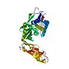
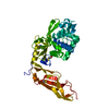
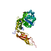
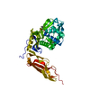
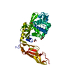
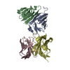
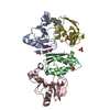
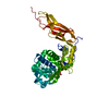
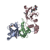
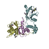
| Title | CRYSTAL STRUCTURE OF A DEACYLATION-DEFECTIVE MUTANT OF PENICILLIN-BINDING PROTEIN 5 MODIFIED BY MERCURY | ||||||
 Components Components | Penicillin-binding protein 5 | ||||||
 Keywords Keywords |  HYDROLASE / PEPTIDOGLYCAN SYNTHESIS / PENICLLIN-BINDING PROTEIN / DD-CARBOXYPEPTIDASE HYDROLASE / PEPTIDOGLYCAN SYNTHESIS / PENICLLIN-BINDING PROTEIN / DD-CARBOXYPEPTIDASE | ||||||
| Function / homology |  Function and homology information Function and homology informationpeptidoglycan metabolic process / serine-type D-Ala-D-Ala carboxypeptidase / serine-type D-Ala-D-Ala carboxypeptidase activity / penicillin binding / peptidoglycan biosynthetic process / carboxypeptidase activity / cell wall organization / beta-lactamase activity / beta-lactamase / outer membrane-bounded periplasmic space ...peptidoglycan metabolic process / serine-type D-Ala-D-Ala carboxypeptidase / serine-type D-Ala-D-Ala carboxypeptidase activity / penicillin binding / peptidoglycan biosynthetic process / carboxypeptidase activity / cell wall organization / beta-lactamase activity / beta-lactamase / outer membrane-bounded periplasmic space / regulation of cell shape / protein homodimerization activity / proteolysis / plasma membraneSimilarity search - Function | ||||||
| Biological species |  Escherichia coli (E. coli) Escherichia coli (E. coli) | ||||||
| Method | X-RAY DIFFRACTION / REFINEMENT / Resolution: 2.5 Å | ||||||
 Authors Authors | Nicola, G. / Nicholas, R.A. / Davies, C. | ||||||
 Citation Citation | Journal: Biochem.J. / Year: 2005 Title: A large displacement of the SXN motif of Cys115-modified penicillin-binding protein 5 from Escherichia coli. Authors: Nicola, G. / Fedarovich, A. / Nicholas, R.A. / Davies, C. #1: Journal: J.Biol.Chem. / Year: 2001Title: Crystal Structure of a Deacylation-Defective Mutant of Penicllin-Binding Protein at 2.3 A Resolution Authors: Davies, C. / White, S.W. / Nicholas, R.A. #2: Journal: J.Biol.Chem. / Year: 2003Title: Crystal Structure of Wild-Type Penicillin-Binding Protein 5 from Escherichia Coli: Implications for Deacylation of the Acyl-Enzyme Complex Authors: Nicholas, R.A. / Krings, S. / Tomberg, J. / Nicola, G. / Davies, C. | ||||||
| History |
| ||||||
| Remark 999 | SEQUENCE THIS IS A SOLUBLE CONSTRUCT OF A MUTANT PBP 5, TERMED SPBP 5'. TO PRODUCE SPBP5 CODONS ...SEQUENCE THIS IS A SOLUBLE CONSTRUCT OF A MUTANT PBP 5, TERMED SPBP 5'. TO PRODUCE SPBP5 CODONS CORRESPONDING TO THE LAST 17 AMINO ACID RESIDUES WERE REMOVED BUT AN ADDITIONAL SIX AMINO ACIDS (GDPVID) WERE ADDED DUE TO READ THROUGH TO THE STOP CODON. NONE OF THESE NON-NATIVE RESIDUES ARE VISIBLE IN THE ELECTRON DENSITY MAP. THE FIRST 29 AMINO ACIDS OF THE PROTEIN ENCODED BY THE OPEN READING FRAME REPRESENT THE SIGNAL SEQUENCE, WHICH IS REMOVED DURING MATURATION AND TRANSPORT TO THE PERIPLASMIC SPACE. THESE RESIDUES ARE NOT PRESENT IN THIS CONSTRUCT. |
- Structure visualization
| Structure viewer | Molecule: MolmilJmol/JSmol |
|---|
- Downloads & links
-Download
| PDBx/mmCIF format | 1sdn.cif.gz | 81.3 KB | Display | PDBx/mmCIF format |
|---|---|---|---|---|
| PDB format | pdb1sdn.ent.gz | 60 KB | Display | PDB format |
| PDBx/mmJSON format | 1sdn.json.gz | Tree view | PDBx/mmJSON format | |
| Others |  Other downloads Other downloads |
-Validation report
| Arichive directory | https://data.pdbj.org/pub/pdb/validation_reports/sd/1sdnftp://data.pdbj.org/pub/pdb/validation_reports/sd/1sdn | HTTPS FTP |
|---|
-Related structure data
| Related structure data | 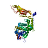 1nzuC 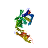 1hd8S C: citing same article ( S: Starting model for refinement |
|---|---|
| Similar structure data |

-Links
 PDBj
PDBj


- Assembly
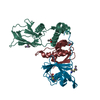
| Deposited unit | 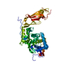
| ||||||||
|---|---|---|---|---|---|---|---|---|---|
| 1 |
| ||||||||
| Unit cell |
|
-Components
| #1: Protein | Mass: 39899.152 Da / Num. of mol.: 1 / Mutation: G105D Source method: isolated from a genetically manipulated source Source: (gene. exp.) Escherichia coli (E. coli) / Gene: DACA, PFV, B0632, C0722, Z0777, ECS0670 / Plasmid: PBR322 / Production host: Escherichia coli (E. coli) / Strain (production host): MC1061References: UniProt: P04287, UniProt: P0AEB2*PLUS, serine-type D-Ala-D-Ala carboxypeptidase |
|---|---|
| #2: Chemical | ChemComp-HG / Mercury (element)  Mass: 200.590 Da / Num. of mol.: 1 / Source method: obtained synthetically / Formula: Hg Mass: 200.590 Da / Num. of mol.: 1 / Source method: obtained synthetically / Formula: Hg |
| #3: Water | ChemComp-HOH / Water Mass: 18.015 Da / Num. of mol.: 46 / Source method: isolated from a natural source / Formula: H2O Mass: 18.015 Da / Num. of mol.: 46 / Source method: isolated from a natural source / Formula: H2O |
-Experimental details
-Experiment
| Experiment | Method: X-RAY DIFFRACTION / Number of used crystals: 1 |
|---|
- Sample preparation
| Crystal | Density Matthews: 2.77 Å3/Da / Density % sol: 55.32 % |
|---|---|
| Crystal grow | Temperature: 294 K / Method: vapor diffusion, hanging drop / pH: 5.5 Details: 24% (w/v) polyethylene glycol 8000, 50mM sodium citrate, 100mM magnesium acetate, pH 5.5, VAPOR DIFFUSION, HANGING DROP, temperature 294K |
-Data collection
| Diffraction | Mean temperature: 294 K |
|---|---|
| Diffraction source | Source: ROTATING ANODE / Type: ENRAF-NONIUS FR591 / Wavelength: 1.5418 Å |
| Detector | Type: MAC Science DIP-2000H / Detector: IMAGE PLATE / Date: Aug 20, 1998 / Details: mirrors |
| Radiation | Protocol: SINGLE WAVELENGTH / Monochromatic (M) / Laue (L): M / Scattering type: x-ray |
| Radiation wavelength | Wavelength: 1.5418 Å / Relative weight: 1 |
| Reflection | Resolution: 2.5→25.4 Å / Num. all: 13482 / Num. obs: 13482 / % possible obs: 95.6 % / Observed criterion σ(F): 0 / Observed criterion σ(I): 0 / Redundancy: 2.53 % / Biso Wilson estimate: 30.1 Å2 / Rmerge(I) obs: 0.074 / Net I/σ(I): 11.2 |
| Reflection shell | Resolution: 2.5→2.59 Å / Rmerge(I) obs: 0.203 / Mean I/σ(I) obs: 4.4 / % possible all: 88.7 |
- Processing
| Software |
| ||||||||||||||||||||||||||||||||||||||||||||||||||||||||||||||||||||||
|---|---|---|---|---|---|---|---|---|---|---|---|---|---|---|---|---|---|---|---|---|---|---|---|---|---|---|---|---|---|---|---|---|---|---|---|---|---|---|---|---|---|---|---|---|---|---|---|---|---|---|---|---|---|---|---|---|---|---|---|---|---|---|---|---|---|---|---|---|---|---|---|
| Refinement | Method to determine structure: REFINEMENT Starting model: PDB ENTRY 1HD8 Resolution: 2.5→15 Å / Cor.coef. Fo:Fc: 0.948 / Cor.coef. Fo:Fc free: 0.901 / SU B: 8.1 / SU ML: 0.182 / Cross valid method: THROUGHOUT / σ(F): 0 / ESU R: 0.494 / ESU R Free: 0.282 / Stereochemistry target values: MAXIMUM LIKELIHOOD
| ||||||||||||||||||||||||||||||||||||||||||||||||||||||||||||||||||||||
| Solvent computation | Ion probe radii: 0.8 Å / Shrinkage radii: 0.8 Å / VDW probe radii: 1.4 Å / Solvent model: BABINET MODEL WITH MASK | ||||||||||||||||||||||||||||||||||||||||||||||||||||||||||||||||||||||
| Displacement parameters | Biso mean: 30.141 Å2
| ||||||||||||||||||||||||||||||||||||||||||||||||||||||||||||||||||||||
| Refinement step | Cycle: LAST / Resolution: 2.5→15 Å
| ||||||||||||||||||||||||||||||||||||||||||||||||||||||||||||||||||||||
| Refine LS restraints |
| ||||||||||||||||||||||||||||||||||||||||||||||||||||||||||||||||||||||
| LS refinement shell | Resolution: 2.5→2.563 Å / Total num. of bins used: 20 /
|
