 Movie
Movie Controller

[English] 日本語
 Yorodumi
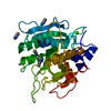
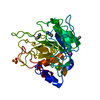
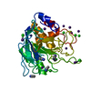
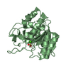
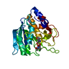
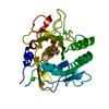
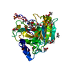
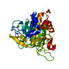
- PDB-1sbc: THE REFINED CRYSTAL STRUCTURE OF SUBTILISIN CARLSBERG AT 2.5 ANGS... -
+ Open data

- Basic information
| Entry | Database: PDB / ID: 1sbc | ||||||
|---|---|---|---|---|---|---|---|
| Title | THE REFINED CRYSTAL STRUCTURE OF SUBTILISIN CARLSBERG AT 2.5 ANGSTROMS RESOLUTION | ||||||
 Components Components | SUBTILISIN CARLSBERG | ||||||
 Keywords Keywords |  SERINE PROTEINASE SERINE PROTEINASE | ||||||
| Function / homology |  Function and homology informationsubtilisin / serine-type endopeptidase activity / proteolysis / extracellular region / metal ion binding Function and homology informationsubtilisin / serine-type endopeptidase activity / proteolysis / extracellular region / metal ion bindingSimilarity search - Function | ||||||
| Biological species |  Bacillus subtilis (bacteria) Bacillus subtilis (bacteria) | ||||||
| Method | X-RAY DIFFRACTION / Resolution: 2.5 Å | ||||||
 Authors Authors | Neidhart, D.J. / Petsko, G.A. | ||||||
 Citation Citation | Journal: Protein Eng. / Year: 1988 Title: The refined crystal structure of subtilisin Carlsberg at 2.5 A resolution. Authors: Neidhart, D.J. / Petsko, G.A. #1: Journal: J.Mol.Biol. / Year: 1976Title: The Structure of Subtilopeptidase A. I. X-Ray Crystallographic Data Authors: Petsko, G.A. / Tsernoglou, D. | ||||||
| History |
|
- Structure visualization
| Structure viewer | Molecule: MolmilJmol/JSmol |
|---|
- Downloads & links
-Download
| PDBx/mmCIF format | 1sbc.cif.gz | 59.4 KB | Display | PDBx/mmCIF format |
|---|---|---|---|---|
| PDB format | pdb1sbc.ent.gz | 43.2 KB | Display | PDB format |
| PDBx/mmJSON format | 1sbc.json.gz | Tree view | PDBx/mmJSON format | |
| Others |  Other downloads Other downloads |
-Validation report
| Arichive directory | https://data.pdbj.org/pub/pdb/validation_reports/sb/1sbcftp://data.pdbj.org/pub/pdb/validation_reports/sb/1sbc | HTTPS FTP |
|---|
-Related structure data
| Similar structure data |
|---|

-Links
 PDBj
PDBj

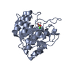
- Assembly
| Deposited unit | 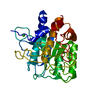
| ||||||||
|---|---|---|---|---|---|---|---|---|---|
| 1 |
| ||||||||
| Unit cell |
| ||||||||
| Atom site foot note | 1: RESIDUE PRO 168 IS A CIS PROLINE. 2: THE DENSITY IS WEAK FOR RESIDUES 54 AND 55 INDICATING POSSIBLE DISORDER. |
-Components
| #1: Protein | Mass: 27306.199 Da / Num. of mol.: 1 Source method: isolated from a genetically manipulated source Source: (gene. exp.) Bacillus subtilis (bacteria) / References: UniProt: P00780, subtilisin |
|---|---|
| #2: Chemical | ChemComp-CA /   Mass: 40.078 Da / Num. of mol.: 1 / Source method: obtained synthetically / Formula: Ca Mass: 40.078 Da / Num. of mol.: 1 / Source method: obtained synthetically / Formula: Ca |
-Experimental details
-Experiment
| Experiment | Method: X-RAY DIFFRACTION |
|---|
- Sample preparation
| Crystal | Density Matthews: 2.07 Å3/Da / Density % sol: 40.66 % | |||||||||||||||
|---|---|---|---|---|---|---|---|---|---|---|---|---|---|---|---|---|
| Crystal | *PLUS Density % sol: 42 % | |||||||||||||||
| Crystal grow | *PLUS pH: 5.6 / Method: batch method / Details: Petsko, G.A., (1976) J.Mol.Biol., 106, 453. | |||||||||||||||
| Components of the solutions | *PLUS
|
- Processing
| Software | Name: PROLSQ / Classification: refinement | ||||||||||||||||||||||||||||||||||||||||||||||||||||||||||||||||||||||||||||||||||||
|---|---|---|---|---|---|---|---|---|---|---|---|---|---|---|---|---|---|---|---|---|---|---|---|---|---|---|---|---|---|---|---|---|---|---|---|---|---|---|---|---|---|---|---|---|---|---|---|---|---|---|---|---|---|---|---|---|---|---|---|---|---|---|---|---|---|---|---|---|---|---|---|---|---|---|---|---|---|---|---|---|---|---|---|---|---|
| Refinement | Rfactor obs: 0.206 / Highest resolution: 2.5 Å Details: THE DENSITY IS WEAK FOR RESIDUES 54 AND 55 INDICATING POSSIBLE DISORDER. | ||||||||||||||||||||||||||||||||||||||||||||||||||||||||||||||||||||||||||||||||||||
| Refinement step | Cycle: LAST / Highest resolution: 2.5 Å
| ||||||||||||||||||||||||||||||||||||||||||||||||||||||||||||||||||||||||||||||||||||
| Refine LS restraints |
| ||||||||||||||||||||||||||||||||||||||||||||||||||||||||||||||||||||||||||||||||||||
| Software | *PLUS Name: PROLSQ / Classification: refinement | ||||||||||||||||||||||||||||||||||||||||||||||||||||||||||||||||||||||||||||||||||||
| Refinement | *PLUS Lowest resolution: 5 Å / σ(F): 1 / Rfactor obs: 0.206 | ||||||||||||||||||||||||||||||||||||||||||||||||||||||||||||||||||||||||||||||||||||
| Solvent computation | *PLUS | ||||||||||||||||||||||||||||||||||||||||||||||||||||||||||||||||||||||||||||||||||||
| Displacement parameters | *PLUS |
