 Movie
Movie Controller


+ Open data

- Basic information
| Entry | Database: PDB / ID: 1s95 | ||||||
|---|---|---|---|---|---|---|---|
| Title | Structure of serine/threonine protein phosphatase 5 | ||||||
 Components Components | Serine/threonine protein phosphatase 5 Serine/threonine-specific protein kinase Serine/threonine-specific protein kinase | ||||||
 Keywords Keywords | HYDROLASE / protein phosphatase / PPPase / PP5 / phosphate anion / metal ion / metallophosphoesterase | ||||||
| Function / homology |  Function and homology information Function and homology informationresponse to arachidonic acid / peptidyl-serine dephosphorylation / peptidyl-threonine dephosphorylation / response to morphine / protein folding chaperone complex / myosin phosphatase activity / protein serine/threonine phosphatase activity / protein-serine/threonine phosphatase / phosphatase activity / phosphoprotein phosphatase activity ...response to arachidonic acid / peptidyl-serine dephosphorylation / peptidyl-threonine dephosphorylation / response to morphine / protein folding chaperone complex / myosin phosphatase activity / protein serine/threonine phosphatase activity / protein-serine/threonine phosphatase / phosphatase activity / phosphoprotein phosphatase activity / ESR-mediated signaling / protein dephosphorylation / ADP binding / response to lead ion / Hsp90 protein binding / tau protein binding / Negative regulation of MAPK pathway / double-strand break repair / MAPK cascade / mitotic cell cycle / Recruitment and ATM-mediated phosphorylation of repair and signaling proteins at DNA double strand breaks / positive regulation of canonical NF-kappaB signal transduction / intracellular membrane-bounded organelle / DNA-templated transcription / lipid binding / protein-containing complex / RNA binding / nucleoplasm / ATP binding / identical protein binding / metal ion binding / nucleus / plasma membrane / cytosolSimilarity search - Function | ||||||
| Biological species |  Homo sapiens (human) Homo sapiens (human) | ||||||
| Method | X-RAY DIFFRACTION / SYNCHROTRON / MOLECULAR REPLACEMENT / Resolution: 1.6 Å | ||||||
 Authors Authors | Swingle, M.R. / Honkanen, R.E. / Ciszak, E.M. | ||||||
 Citation Citation | Journal: J.Biol.Chem. / Year: 2004 Title: Structural basis for the catalytic activity of human serine/threonine protein phosphatase-5. Authors: Swingle, M.R. / Honkanen, R.E. / Ciszak, E.M. | ||||||
| History |
|
- Structure visualization
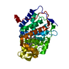
| Structure viewer | Molecule: MolmilJmol/JSmol |
|---|
- Downloads & links
-Download
| PDBx/mmCIF format | 1s95.cif.gz | 153.9 KB | Display | PDBx/mmCIF format |
|---|---|---|---|---|
| PDB format | pdb1s95.ent.gz | 118.8 KB | Display | PDB format |
| PDBx/mmJSON format | 1s95.json.gz | Tree view | PDBx/mmJSON format | |
| Others |  Other downloads Other downloads |
-Validation report
| Arichive directory | https://data.pdbj.org/pub/pdb/validation_reports/s9/1s95ftp://data.pdbj.org/pub/pdb/validation_reports/s9/1s95 | HTTPS FTP |
|---|
-Related structure data


-Links
 PDBj
PDBj


- Assembly
| Deposited unit | 
| ||||||||
|---|---|---|---|---|---|---|---|---|---|
| 1 | 
| ||||||||
| 2 | 
| ||||||||
| 3 |
| ||||||||
| Unit cell |
|
-Components
| #1: Protein | Serine/threonine-specific protein kinase / PP5 / Protein phosphatase T / PP-T / PPT Mass: 37887.020 Da / Num. of mol.: 2 / Fragment: catalytic domain Source method: isolated from a genetically manipulated source Source: (gene. exp.) Homo sapiens (human) / Gene: PPP5C, PPP5 / Plasmid: pMalc2E / Species (production host): Escherichia coli / Production host:  Escherichia coli BL21 (bacteria) / Strain (production host): BL21 Escherichia coli BL21 (bacteria) / Strain (production host): BL21References: UniProt: P53041, protein-serine/threonine phosphatase #2: Chemical | ChemComp-MN /   Mass: 54.938 Da / Num. of mol.: 4 / Source method: obtained synthetically / Formula: Mn Mass: 54.938 Da / Num. of mol.: 4 / Source method: obtained synthetically / Formula: Mn#3: Chemical | Phosphate  Mass: 94.971 Da / Num. of mol.: 2 / Source method: obtained synthetically / Formula: PO4 Mass: 94.971 Da / Num. of mol.: 2 / Source method: obtained synthetically / Formula: PO4#4: Chemical | 2-Methyl-2,4-pentanediol  Mass: 118.174 Da / Num. of mol.: 2 / Source method: obtained synthetically / Formula: C6H14O2 / Comment: precipitant*YM Mass: 118.174 Da / Num. of mol.: 2 / Source method: obtained synthetically / Formula: C6H14O2 / Comment: precipitant*YM#5: Water | ChemComp-HOH / | Water Mass: 18.015 Da / Num. of mol.: 463 / Source method: isolated from a natural source / Formula: H2O Mass: 18.015 Da / Num. of mol.: 463 / Source method: isolated from a natural source / Formula: H2O |
|---|
-Experimental details
-Experiment
| Experiment | Method: X-RAY DIFFRACTION / Number of used crystals: 1 |
|---|
- Sample preparation
| Crystal | Density Matthews: 2.45 Å3/Da / Density % sol: 30 % |
|---|---|
| Crystal grow | Temperature: 298 K / Method: vapor diffusion, hanging drop / pH: 7.5 Details: 2-methyl,2,4-pentanediol, pH 7.5, VAPOR DIFFUSION, HANGING DROP, temperature 298K |
-Data collection
| Diffraction | Mean temperature: 100 K |
|---|---|
| Diffraction source | Source: SYNCHROTRON / Site: APS  / Beamline: 22-ID / Wavelength: 1 Å / Beamline: 22-ID / Wavelength: 1 Å |
| Detector | Type: MARRESEARCH / Detector: CCD / Date: Nov 10, 2002 |
| Radiation | Protocol: SINGLE WAVELENGTH / Monochromatic (M) / Laue (L): M / Scattering type: x-ray |
| Radiation wavelength | Wavelength: 1 Å / Relative weight: 1 |
| Reflection | Resolution: 1.47→40 Å / Num. all: 70964 / Num. obs: 70964 / % possible obs: 70 % / Observed criterion σ(F): 2 / Observed criterion σ(I): 0 / Redundancy: 2.9 % / Rmerge(I) obs: 0.064 / Rsym value: 0.064 / Net I/σ(I): 8.9 |
| Reflection shell | Resolution: 1.47→1.59 Å / Redundancy: 1 % / % possible all: 13 |
- Processing
| Software |
| |||||||||||||||||||||||||||||||||||||||||||||||||||||||||||||||||||||||||||||||||||||||||||||||||||||||||||||||||||||||||||||
|---|---|---|---|---|---|---|---|---|---|---|---|---|---|---|---|---|---|---|---|---|---|---|---|---|---|---|---|---|---|---|---|---|---|---|---|---|---|---|---|---|---|---|---|---|---|---|---|---|---|---|---|---|---|---|---|---|---|---|---|---|---|---|---|---|---|---|---|---|---|---|---|---|---|---|---|---|---|---|---|---|---|---|---|---|---|---|---|---|---|---|---|---|---|---|---|---|---|---|---|---|---|---|---|---|---|---|---|---|---|---|---|---|---|---|---|---|---|---|---|---|---|---|---|---|---|---|
| Refinement | Method to determine structure: MOLECULAR REPLACEMENT Starting model: Partial homology model derived from 1IT6, 1AUJ, 1G5B Resolution: 1.6→40 Å / Cor.coef. Fo:Fc: 0.967 / Cor.coef. Fo:Fc free: 0.951 / SU B: 4.112 / SU ML: 0.074 / TLS residual ADP flag: LIKELY RESIDUAL / Cross valid method: THROUGHOUT / σ(F): 2 / σ(I): 0 / ESU R: 0.106 / ESU R Free: 0.105 / Stereochemistry target values: MAXIMUM LIKELIHOOD
| |||||||||||||||||||||||||||||||||||||||||||||||||||||||||||||||||||||||||||||||||||||||||||||||||||||||||||||||||||||||||||||
| Solvent computation | Ion probe radii: 0.8 Å / Shrinkage radii: 0.8 Å / VDW probe radii: 1.2 Å / Solvent model: BABINET MODEL WITH MASK | |||||||||||||||||||||||||||||||||||||||||||||||||||||||||||||||||||||||||||||||||||||||||||||||||||||||||||||||||||||||||||||
| Displacement parameters | Biso mean: 23.31 Å2
| |||||||||||||||||||||||||||||||||||||||||||||||||||||||||||||||||||||||||||||||||||||||||||||||||||||||||||||||||||||||||||||
| Refine analyze |
| |||||||||||||||||||||||||||||||||||||||||||||||||||||||||||||||||||||||||||||||||||||||||||||||||||||||||||||||||||||||||||||
| Refinement step | Cycle: LAST / Resolution: 1.6→40 Å
| |||||||||||||||||||||||||||||||||||||||||||||||||||||||||||||||||||||||||||||||||||||||||||||||||||||||||||||||||||||||||||||
| Refine LS restraints |
| |||||||||||||||||||||||||||||||||||||||||||||||||||||||||||||||||||||||||||||||||||||||||||||||||||||||||||||||||||||||||||||
| LS refinement shell | Resolution: 1.6→1.68 Å / Total num. of bins used: 10 /
| |||||||||||||||||||||||||||||||||||||||||||||||||||||||||||||||||||||||||||||||||||||||||||||||||||||||||||||||||||||||||||||
| Refinement TLS params. | Method: refined / Refine-ID: X-RAY DIFFRACTION
| |||||||||||||||||||||||||||||||||||||||||||||||||||||||||||||||||||||||||||||||||||||||||||||||||||||||||||||||||||||||||||||
| Refinement TLS group |
|
