 Movie
Movie Controller

[English] 日本語
 Yorodumi
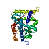
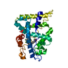
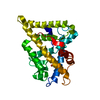
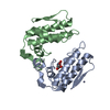
- PDB-1s6b: X-ray Crystal Structure of a Complex Formed Between Two Homologou... -
+ Open data

- Basic information
| Entry | Database: PDB / ID: 1s6b | |||||||||
|---|---|---|---|---|---|---|---|---|---|---|
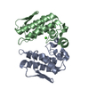
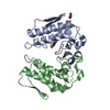
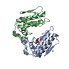
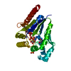
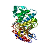
| Title | X-ray Crystal Structure of a Complex Formed Between Two Homologous Isoforms of Phospholipase A2 from Naja naja sagittifera: Principle of Molecular Association and Inactivation | |||||||||
 Components Components | (Phospholipase A2 isoform ...) x 2 | |||||||||
 Keywords Keywords |  HYDROLASE / Phospholipase A2 / molecular conformation / enzyme activity HYDROLASE / Phospholipase A2 / molecular conformation / enzyme activity | |||||||||
| Function / homology |  Function and homology informationphospholipase A2 / phospholipase A2 activity / arachidonic acid secretion / phospholipid metabolic process / lipid catabolic process / calcium ion binding / extracellular region Function and homology informationphospholipase A2 / phospholipase A2 activity / arachidonic acid secretion / phospholipid metabolic process / lipid catabolic process / calcium ion binding / extracellular regionSimilarity search - Function | |||||||||
| Biological species |  Naja sagittifera (cobra) Naja sagittifera (cobra) | |||||||||
| Method | X-RAY DIFFRACTION / SYNCHROTRON / MOLECULAR REPLACEMENT / Resolution: 1.6 Å | |||||||||
 Authors Authors | Jabeen, T. / Sharma, S. / Singh, R.K. / Kaur, P. / Singh, T.P. | |||||||||
 Citation Citation | Journal: Proteins / Year: 2005 Title: Crystal structure of a calcium-induced dimer of two isoforms of cobra phospholipase A2 at 1.6 A resolution. Authors: Jabeen, T. / Sharma, S. / Singh, N. / Singh, R.K. / Kaur, P. / Perbandt, M. / Betzel, C.h. / Srinivasan, A. / Singh, T.P. #1: Journal: To be PublishedTitle: Crystal Structure of an Acidic Phospholipase A2 from Naja Naja Sagittifera at 1.5 A Resolution Authors: Singh, R.K. / Sharma, S. / Jabeen, T. / Kaur, P. / Singh, T.P. | |||||||||
| History |
|
- Structure visualization
| Structure viewer | Molecule: MolmilJmol/JSmol |
|---|
- Downloads & links
-Download
| PDBx/mmCIF format | 1s6b.cif.gz | 69.8 KB | Display | PDBx/mmCIF format |
|---|---|---|---|---|
| PDB format | pdb1s6b.ent.gz | 49.6 KB | Display | PDB format |
| PDBx/mmJSON format | 1s6b.json.gz | Tree view | PDBx/mmJSON format | |
| Others |  Other downloads Other downloads |
-Validation report
| Arichive directory | https://data.pdbj.org/pub/pdb/validation_reports/s6/1s6bftp://data.pdbj.org/pub/pdb/validation_reports/s6/1s6b | HTTPS FTP |
|---|
-Related structure data
| Related structure data | 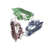 1pshS S: Starting model for refinement |
|---|---|
| Similar structure data |

-Links
 PDBj
PDBj


- Assembly
| Deposited unit | 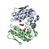
| ||||||||
|---|---|---|---|---|---|---|---|---|---|
| 1 |
| ||||||||
| Unit cell |
|
-Components
-Phospholipase A2 isoform ... , 2 types, 2 molecules AB
| #1: Protein | Mass: 13220.687 Da / Num. of mol.: 1 / Source method: isolated from a natural source / Source: (natural) Naja sagittifera (cobra) / Secretion: venom / References: UniProt: P60043, phospholipase A2 |
|---|---|
| #2: Protein | Mass: 13288.681 Da / Num. of mol.: 1 / Source method: isolated from a natural source / Source: (natural) Naja sagittifera (cobra) / Secretion: venom / References: UniProt: P60044, phospholipase A2 |
-Non-polymers , 4 types, 360 molecules 


| #3: Chemical |  Mass: 40.078 Da / Num. of mol.: 2 / Source method: obtained synthetically / Formula: Ca Mass: 40.078 Da / Num. of mol.: 2 / Source method: obtained synthetically / Formula: Ca#4: Chemical | Phosphate Mass: 94.971 Da / Num. of mol.: 2 / Source method: obtained synthetically / Formula: PO4 Mass: 94.971 Da / Num. of mol.: 2 / Source method: obtained synthetically / Formula: PO4#5: Chemical | Acetic acid Mass: 60.052 Da / Num. of mol.: 2 / Source method: obtained synthetically / Formula: C2H4O2 Mass: 60.052 Da / Num. of mol.: 2 / Source method: obtained synthetically / Formula: C2H4O2#6: Water | ChemComp-HOH / | WaterMass: 18.015 Da / Num. of mol.: 354 / Source method: isolated from a natural source / Formula: H2O |
|---|
-Experimental details
-Experiment
| Experiment | Method: X-RAY DIFFRACTION / Number of used crystals: 1 |
|---|
- Sample preparation
| Crystal | Density Matthews: 2.3 Å3/Da / Density % sol: 46 % | |||||||||||||||||||||||||
|---|---|---|---|---|---|---|---|---|---|---|---|---|---|---|---|---|---|---|---|---|---|---|---|---|---|---|
| Crystal grow | Temperature: 298 K / Method: vapor diffusion, sitting drop / pH: 6 Details: calcium chloride, tris, pH 6.00, VAPOR DIFFUSION, SITTING DROP, temperature 298K | |||||||||||||||||||||||||
| Crystal grow | *PLUS Method: vapor diffusion, sitting drop | |||||||||||||||||||||||||
| Components of the solutions | *PLUS
|
-Data collection
| Diffraction | Mean temperature: 200 K | |||||||||
|---|---|---|---|---|---|---|---|---|---|---|
| Diffraction source | Source: SYNCHROTRON / Site: EMBL/DESY, HAMBURG  / Beamline: X11 / Wavelength: 0.802 / Wavelength: 0.91 Å / Beamline: X11 / Wavelength: 0.802 / Wavelength: 0.91 Å | |||||||||
| Detector | Type: MARRESEARCH / Detector: CCD / Date: Sep 11, 2001 / Details: MIRRORS | |||||||||
| Radiation | Monochromator: MIRROR / Protocol: SINGLE WAVELENGTH / Monochromatic (M) / Laue (L): M / Scattering type: x-ray | |||||||||
| Radiation wavelength |
| |||||||||
| Reflection | Resolution: 1.6→20 Å / Num. obs: 27946 / % possible obs: 98.6 % / Observed criterion σ(I): -3 / Redundancy: 17.9 % / Biso Wilson estimate: 29.1 Å2 / Rsym value: 0.043 / Net I/σ(I): 26 | |||||||||
| Reflection shell | Resolution: 1.6→1.68 Å / Mean I/σ(I) obs: 3.02 / Rsym value: 0.329 / % possible all: 97.3 | |||||||||
| Reflection | *PLUS Highest resolution: 1.6 Å / Lowest resolution: 20 Å / Num. measured all: 500751 / Rmerge(I) obs: 0.043 | |||||||||
| Reflection shell | *PLUS % possible obs: 97.3 % / Rmerge(I) obs: 0.329 / Mean I/σ(I) obs: 3 |
- Processing
| Software |
| |||||||||||||||||||||||||||||||||||||||||||||||||||||||||||||||
|---|---|---|---|---|---|---|---|---|---|---|---|---|---|---|---|---|---|---|---|---|---|---|---|---|---|---|---|---|---|---|---|---|---|---|---|---|---|---|---|---|---|---|---|---|---|---|---|---|---|---|---|---|---|---|---|---|---|---|---|---|---|---|---|---|
| Refinement | Method to determine structure: MOLECULAR REPLACEMENT Starting model: PDB ENTRY 1PSH Resolution: 1.6→20 Å / Cross valid method: THROUGHOUT / σ(F): 0 / Stereochemistry target values: Engh & Huber / Details: USED CONJUGATE DIRECTION METHOD
| |||||||||||||||||||||||||||||||||||||||||||||||||||||||||||||||
| Displacement parameters | Biso mean: 30.8 Å2 | |||||||||||||||||||||||||||||||||||||||||||||||||||||||||||||||
| Refinement step | Cycle: LAST / Resolution: 1.6→20 Å
| |||||||||||||||||||||||||||||||||||||||||||||||||||||||||||||||
| Refine LS restraints |
| |||||||||||||||||||||||||||||||||||||||||||||||||||||||||||||||
| Refinement | *PLUS Highest resolution: 1.6 Å / Lowest resolution: 20 Å / Rfactor Rwork: 0.21 | |||||||||||||||||||||||||||||||||||||||||||||||||||||||||||||||
| Solvent computation | *PLUS | |||||||||||||||||||||||||||||||||||||||||||||||||||||||||||||||
| Displacement parameters | *PLUS | |||||||||||||||||||||||||||||||||||||||||||||||||||||||||||||||
| Refine LS restraints | *PLUS
|
