 Movie
Movie Controller

[English] 日本語
 Yorodumi
- PDB-1rtd: STRUCTURE OF A CATALYTIC COMPLEX OF HIV-1 REVERSE TRANSCRIPTASE: ... -
+ Open data

- Basic information
| Entry | Database: PDB / ID: 1rtd | ||||||
|---|---|---|---|---|---|---|---|
| Title | STRUCTURE OF A CATALYTIC COMPLEX OF HIV-1 REVERSE TRANSCRIPTASE: IMPLICATIONS FOR NUCLEOSIDE ANALOG DRUG RESISTANCE | ||||||
 Components Components |
| ||||||
 Keywords Keywords | TRANSFERASE/DNA / COMPLEX(NUCLEOTIDYLTRANSFERASE /  DNA / DNTP) / PROTEIN/DNA / TRANSFERASE-DNA COMPLEX DNA / DNTP) / PROTEIN/DNA / TRANSFERASE-DNA COMPLEX | ||||||
| Function / homology |  Function and homology informationintegrase activity / Integration of viral DNA into host genomic DNA / Autointegration results in viral DNA circles / Minus-strand DNA synthesis / Plus-strand DNA synthesis / Uncoating of the HIV Virion / 2-LTR circle formation / Vpr-mediated nuclear import of PICs / Early Phase of HIV Life Cycle / Integration of provirus ...integrase activity / Integration of viral DNA into host genomic DNA / Autointegration results in viral DNA circles / Minus-strand DNA synthesis / Plus-strand DNA synthesis / Uncoating of the HIV Virion / 2-LTR circle formation / Vpr-mediated nuclear import of PICs / Early Phase of HIV Life Cycle / Integration of provirus / APOBEC3G mediated resistance to HIV-1 infection / Binding and entry of HIV virion / viral life cycle / Assembly Of The HIV Virion / HIV-1 retropepsin / : / Budding and maturation of HIV virion / retroviral ribonuclease H / exoribonuclease H / : / exoribonuclease H activity / protein processing / host multivesicular body / DNA integration / RNA-directed DNA polymerase / viral genome integration into host DNA / viral penetration into host nucleus / establishment of integrated proviral latency / RNA-directed DNA polymerase activity / Transferases; Transferring phosphorus-containing groups; Nucleotidyltransferases / RNA-DNA hybrid ribonuclease activity / peptidase activity / viral nucleocapsid / DNA recombination / Hydrolases; Acting on ester bonds / DNA-directed DNA polymerase / aspartic-type endopeptidase activity / DNA-directed DNA polymerase activity / symbiont entry into host cell / symbiont-mediated suppression of host gene expression / lipid binding / host cell nucleus / structural molecule activity / host cell plasma membrane / virion membrane / proteolysis / DNA binding / RNA binding / zinc ion binding / membrane / identical protein binding Function and homology informationintegrase activity / Integration of viral DNA into host genomic DNA / Autointegration results in viral DNA circles / Minus-strand DNA synthesis / Plus-strand DNA synthesis / Uncoating of the HIV Virion / 2-LTR circle formation / Vpr-mediated nuclear import of PICs / Early Phase of HIV Life Cycle / Integration of provirus ...integrase activity / Integration of viral DNA into host genomic DNA / Autointegration results in viral DNA circles / Minus-strand DNA synthesis / Plus-strand DNA synthesis / Uncoating of the HIV Virion / 2-LTR circle formation / Vpr-mediated nuclear import of PICs / Early Phase of HIV Life Cycle / Integration of provirus / APOBEC3G mediated resistance to HIV-1 infection / Binding and entry of HIV virion / viral life cycle / Assembly Of The HIV Virion / HIV-1 retropepsin / : / Budding and maturation of HIV virion / retroviral ribonuclease H / exoribonuclease H / : / exoribonuclease H activity / protein processing / host multivesicular body / DNA integration / RNA-directed DNA polymerase / viral genome integration into host DNA / viral penetration into host nucleus / establishment of integrated proviral latency / RNA-directed DNA polymerase activity / Transferases; Transferring phosphorus-containing groups; Nucleotidyltransferases / RNA-DNA hybrid ribonuclease activity / peptidase activity / viral nucleocapsid / DNA recombination / Hydrolases; Acting on ester bonds / DNA-directed DNA polymerase / aspartic-type endopeptidase activity / DNA-directed DNA polymerase activity / symbiont entry into host cell / symbiont-mediated suppression of host gene expression / lipid binding / host cell nucleus / structural molecule activity / host cell plasma membrane / virion membrane / proteolysis / DNA binding / RNA binding / zinc ion binding / membrane / identical protein bindingSimilarity search - Function | ||||||
| Biological species |   Human immunodeficiency virus 1 Human immunodeficiency virus 1 | ||||||
| Method | X-RAY DIFFRACTION / SYNCHROTRON / MOLECULAR REPLACEMENT / Resolution: 3.2 Å | ||||||
 Authors Authors | Chopra, R. / Huang, H. / Verdine, G.L. / Harrison, S.C. | ||||||
 Citation Citation | Journal: Science / Year: 1998 Title: Structure of a covalently trapped catalytic complex of HIV-1 reverse transcriptase: implications for drug resistance. Authors: Huang, H. / Chopra, R. / Verdine, G.L. / Harrison, S.C. | ||||||
| History |
|
- Structure visualization
| Structure viewer | Molecule: MolmilJmol/JSmol |
|---|
- Downloads & links
-Download
| PDBx/mmCIF format | 1rtd.cif.gz | 447.8 KB | Display | PDBx/mmCIF format |
|---|---|---|---|---|
| PDB format | pdb1rtd.ent.gz | 360.9 KB | Display | PDB format |
| PDBx/mmJSON format | 1rtd.json.gz | Tree view | PDBx/mmJSON format | |
| Others |  Other downloads Other downloads |
-Validation report
| Arichive directory | https://data.pdbj.org/pub/pdb/validation_reports/rt/1rtdftp://data.pdbj.org/pub/pdb/validation_reports/rt/1rtd | HTTPS FTP |
|---|
-Related structure data
| Similar structure data |
|---|


-Links
 PDBj
PDBj


- Assembly
| Deposited unit | 
| ||||||||||||||||||||||||
|---|---|---|---|---|---|---|---|---|---|---|---|---|---|---|---|---|---|---|---|---|---|---|---|---|---|
| 1 | 
| ||||||||||||||||||||||||
| 2 | 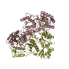
| ||||||||||||||||||||||||
| Unit cell |
| ||||||||||||||||||||||||
| Noncrystallographic symmetry (NCS) | NCS oper:
| ||||||||||||||||||||||||
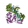
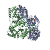
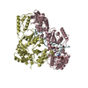
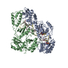
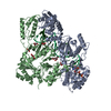
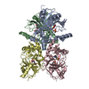
| Details | THE HIV-1 REVERSE TRANSCRIPTASE CONSISTS OF TWO SUBUNITS, P66 (DESIGNATED BY CHAINS A AND C) AND P51 (DESIGNATED BY CHAINS B AND D). THE DNA DUPLEX IS COMPOSED OF TEMPLATE (DESIGNATED CHAIN E) AND PRIMER (DESIGNATED CHAIN F). THE BOUND DEOXY-THYMINE TRIPHOSPHATE HAS CHAIN IDENTIFIER A. IN THIS CRYSTAL FORM THERE ARE TWO REVERSE TRANSCRIPTASE COMPLEXES PER ASYMMETRIC UNIT. ONE MOLECULE WAS SIGNIFICANTLY BETTER ORDERED THAN THE OTHER. COORDINATES FOR BOTH MOLECULES ARE CONTAINED IN THIS ENTRY, AND THE NON-CRYSTALLOGRAPHIC SYMMETRY TRANSFORMATIONS GIVEN BELOW WERE THOSE USED TO GENERATE CHAINS C,D,G, AND H FROM A,B,E, AND F DURING THE COURSE OF REFINEMENT. |
-Components
-DNA chain , 2 types, 4 molecules EGFH
| #1: DNA chain | Mass: 8327.361 Da / Num. of mol.: 2 / Mutation: C2-THIOL TETHER AT TEMPLATE GUANINE 11 / Source method: obtained synthetically #2: DNA chain | Mass: 6416.123 Da / Num. of mol.: 2 / Fragment: PRIMER / Mutation: C2-THIOL TETHER AT TEMPLATE GUANINE 11 / Source method: obtained synthetically |
|---|
-PROTEIN (REVERSE ... , 2 types, 4 molecules ACBD
| #3: Protein | Mass: 63869.148 Da / Num. of mol.: 2 / Fragment: P61 / Mutation: P1K, Q258C, E478Q Source method: isolated from a genetically manipulated source Source: (gene. exp.) Human immunodeficiency virus 1 / Genus: Lentivirus / Strain: BH10 / Gene: POL / Production host:  Escherichia coli (E. coli) / References: UniProt: P03366, RNA-directed DNA polymerase Escherichia coli (E. coli) / References: UniProt: P03366, RNA-directed DNA polymerase#4: Protein | Mass: 51399.047 Da / Num. of mol.: 2 / Fragment: P50 Source method: isolated from a genetically manipulated source Source: (gene. exp.) Human immunodeficiency virus 1 / Genus: Lentivirus / Strain: BH10 / Gene: POL / Production host: Escherichia coli (E. coli) / References: UniProt: P04585, RNA-directed DNA polymerase |
|---|
-Non-polymers , 2 types, 8 molecules 

| #5: Chemical | ChemComp-MG /  Mass: 24.305 Da / Num. of mol.: 6 / Source method: obtained synthetically / Formula: Mg Mass: 24.305 Da / Num. of mol.: 6 / Source method: obtained synthetically / Formula: Mg#6: Chemical | Thymidine triphosphate Mass: 482.168 Da / Num. of mol.: 2 / Source method: obtained synthetically / Formula: C10H17N2O14P3 Mass: 482.168 Da / Num. of mol.: 2 / Source method: obtained synthetically / Formula: C10H17N2O14P3 |
|---|
-Experimental details
-Experiment
| Experiment | Method: X-RAY DIFFRACTION / Number of used crystals: 1 |
|---|
- Sample preparation
| Crystal | Density Matthews: 3.21 Å3/Da / Density % sol: 61.66 % | ||||||||||||||||||||||||||||||||||||||||||||||||||||||||||||||||||||||||
|---|---|---|---|---|---|---|---|---|---|---|---|---|---|---|---|---|---|---|---|---|---|---|---|---|---|---|---|---|---|---|---|---|---|---|---|---|---|---|---|---|---|---|---|---|---|---|---|---|---|---|---|---|---|---|---|---|---|---|---|---|---|---|---|---|---|---|---|---|---|---|---|---|---|
| Crystal grow | pH: 6.5 / Details: pH 6.5 | ||||||||||||||||||||||||||||||||||||||||||||||||||||||||||||||||||||||||
| Crystal grow | *PLUS Temperature: 4 ℃ / Method: vapor diffusion, hanging dropDetails: drop consists of equal volume of protein and reservoir solutions | ||||||||||||||||||||||||||||||||||||||||||||||||||||||||||||||||||||||||
| Components of the solutions | *PLUS
|
-Data collection
| Diffraction | Mean temperature: 110 K |
|---|---|
| Diffraction source | Source: SYNCHROTRON / Site: NSLS  / Beamline: X25 / Wavelength: 1.1 / Beamline: X25 / Wavelength: 1.1 |
| Detector | Type: BRANDEIS - B4 / Detector: CCD |
| Radiation | Protocol: SINGLE WAVELENGTH / Monochromatic (M) / Laue (L): M / Scattering type: x-ray |
| Radiation wavelength | Wavelength: 1.1 Å / Relative weight: 1 |
| Reflection | Resolution: 3.2→18 Å / Num. obs: 54281 / % possible obs: 93.8 % / Observed criterion σ(I): 2 / Redundancy: 4.2 % / Rsym value: 0.103 / Net I/σ(I): 11.2 |
| Reflection shell | Resolution: 3.2→3.3 Å / Redundancy: 1.7 % / Mean I/σ(I) obs: 2.7 / Rsym value: 0.372 / % possible all: 84.3 |
| Reflection | *PLUS Rmerge(I) obs: 0.103 |
| Reflection shell | *PLUS % possible obs: 84.3 % / Num. unique obs: 4802 / Rmerge(I) obs: 0.372 |
- Processing
| Software |
| ||||||||||||||||||||||||||||||||||||||||||||||||||||||||||||
|---|---|---|---|---|---|---|---|---|---|---|---|---|---|---|---|---|---|---|---|---|---|---|---|---|---|---|---|---|---|---|---|---|---|---|---|---|---|---|---|---|---|---|---|---|---|---|---|---|---|---|---|---|---|---|---|---|---|---|---|---|---|
| Refinement | Method to determine structure: MOLECULAR REPLACEMENT / Resolution: 3.2→12 Å / Cross valid method: THROUGHOUT / σ(F): 2
| ||||||||||||||||||||||||||||||||||||||||||||||||||||||||||||
| Refinement step | Cycle: LAST / Resolution: 3.2→12 Å
| ||||||||||||||||||||||||||||||||||||||||||||||||||||||||||||
| Refine LS restraints |
| ||||||||||||||||||||||||||||||||||||||||||||||||||||||||||||
| Refine LS restraints NCS | NCS model details: RESTRAINTS | ||||||||||||||||||||||||||||||||||||||||||||||||||||||||||||
| LS refinement shell | Resolution: 3.2→3.3 Å / Total num. of bins used: 10
| ||||||||||||||||||||||||||||||||||||||||||||||||||||||||||||
| Xplor file |
|
