 Movie
Movie Controller

[English] 日本語
 Yorodumi
- PDB-1rpa: THREE-DIMENSIONAL STRUCTURE OF RAT ACID PHOSPHATASE IN COMPLEX WI... -
+ Open data

- Basic information
| Entry | Database: PDB / ID: 1rpa | |||||||||
|---|---|---|---|---|---|---|---|---|---|---|
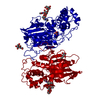
| Title | THREE-DIMENSIONAL STRUCTURE OF RAT ACID PHOSPHATASE IN COMPLEX WITH L(+) TARTRATE | |||||||||
 Components Components | PROSTATIC ACID PHOSPHATASE | |||||||||
 Keywords Keywords | HYDROLASE(PHOSPHORIC MONOESTER) | |||||||||
| Function / homology |  Function and homology information Function and homology informationthiamine phosphate phosphatase activity / positive regulation of adenosine receptor signaling pathway / thiamine metabolic process / Golgi cisterna / adenosine metabolic process / acid phosphatase / regulation of sensory perception of pain / lysophosphatidic acid phosphatase activity / acid phosphatase activity / XMP 5'-nucleosidase activity ...thiamine phosphate phosphatase activity / positive regulation of adenosine receptor signaling pathway / thiamine metabolic process / Golgi cisterna / adenosine metabolic process / acid phosphatase / regulation of sensory perception of pain / lysophosphatidic acid phosphatase activity / acid phosphatase activity / XMP 5'-nucleosidase activity / 5'-nucleotidase / 5'-nucleotidase activity / vesicle membrane / nucleotide metabolic process / choline binding / Neutrophil degranulation / lysosome organization / phosphatase activity / purine nucleobase metabolic process / dephosphorylation / multivesicular body / protein-tyrosine-phosphatase / filopodium / secretory granule / protein tyrosine phosphatase activity / lipid metabolic process / apical part of cell / lysosome / molecular adaptor activity / lysosomal membrane / protein homodimerization activity / extracellular space / membrane / identical protein binding / plasma membraneSimilarity search - Function | |||||||||
| Biological species |  Rattus norvegicus (Norway rat) Rattus norvegicus (Norway rat) | |||||||||
| Method | X-RAY DIFFRACTION / Resolution: 3 Å | |||||||||
 Authors Authors | Lindqvist, Y. / Schneider, G. | |||||||||
 Citation Citation | Journal: J.Biol.Chem. / Year: 1993 Title: Three-dimensional structure of rat acid phosphatase in complex with L(+)-tartrate. Authors: Lindqvist, Y. / Schneider, G. / Vihko, P. #1: Journal: Embo J. / Year: 1993Title: Three-Dimensional Structure of Rat Acid Phosphatase Authors: Schneider, G. / Lindqvist, Y. / Vihko, P. #2: Journal: Proc.Natl.Acad.Sci.USA / Year: 1993Title: Rat Acid Phosphatase: Overexpression of Active, Secreted Enzyme by Recombinant Baculovirus-Infected Insect Cells, Molecular Properties, and Crystallization Authors: Vihko, P. / Kurkela, R. / Porvari, K. / Herrala, A. / Lindfors, A. / Lindqvist, Y. / Schneider, G. | |||||||||
| History |
|
- Structure visualization
| Structure viewer | Molecule: MolmilJmol/JSmol |
|---|
- Downloads & links
-Download
| PDBx/mmCIF format | 1rpa.cif.gz | 71.9 KB | Display | PDBx/mmCIF format |
|---|---|---|---|---|
| PDB format | pdb1rpa.ent.gz | 54.5 KB | Display | PDB format |
| PDBx/mmJSON format | 1rpa.json.gz | Tree view | PDBx/mmJSON format | |
| Others |  Other downloads Other downloads |
-Validation report
| Arichive directory | https://data.pdbj.org/pub/pdb/validation_reports/rp/1rpaftp://data.pdbj.org/pub/pdb/validation_reports/rp/1rpa | HTTPS FTP |
|---|
-Related structure data
| Similar structure data |
|---|


-Links
 PDBj
PDBj


- Assembly
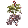
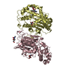
| Deposited unit | 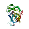
| ||||||||
|---|---|---|---|---|---|---|---|---|---|
| 1 | 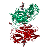
| ||||||||
| Unit cell |
| ||||||||
| Atom site foot note | 1: CIS PROLINE - PRO 125 2: GLN 244 - PRO 245 OMEGA =235.93 PEPTIDE BOND DEVIATES SIGNIFICANTLY FROM TRANS CONFORMATION |
-Components
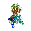
| #1: Protein | Mass: 39691.215 Da / Num. of mol.: 1 Source method: isolated from a genetically manipulated source Source: (gene. exp.) Rattus norvegicus (Norway rat) / References: UniProt: P20646, acid phosphatase |
|---|---|
| #2: Polysaccharide | beta-D-mannopyranose-(1-4)-2-acetamido-2-deoxy-beta-D-glucopyranose-(1-4)-2-acetamido-2-deoxy-beta- ...beta-D-mannopyranose-(1-4)-2-acetamido-2-deoxy-beta-D-glucopyranose-(1-4)-2-acetamido-2-deoxy-beta-D-glucopyranose / Mass: 586.542 Da / Num. of mol.: 1 Source method: isolated from a genetically manipulated source |
| #3: Sugar | ChemComp-NAG / N-Acetylglucosamine  Type: D-saccharide, beta linking / Mass: 221.208 Da / Num. of mol.: 1 Type: D-saccharide, beta linking / Mass: 221.208 Da / Num. of mol.: 1Source method: isolated from a genetically manipulated source Formula: C8H15NO6 |
| #4: Chemical | ChemComp-TAR / Tartaric acid  Mass: 150.087 Da / Num. of mol.: 1 / Source method: obtained synthetically / Formula: C4H6O6 Mass: 150.087 Da / Num. of mol.: 1 / Source method: obtained synthetically / Formula: C4H6O6 |
| Sequence details | SEQUENCE ADVISORY NOTICE DIFFERENCE BETWEEN SWISS-PROT AND PDB SEQUENCE. SWISS-PROT ENTRY NAME: ...SEQUENCE ADVISORY NOTICE DIFFERENCE |
-Experimental details
-Experiment
| Experiment | Method: X-RAY DIFFRACTION |
|---|
- Sample preparation
| Crystal | Density Matthews: 4.42 Å3/Da / Density % sol: 72.14 % | ||||||||||||||||||||
|---|---|---|---|---|---|---|---|---|---|---|---|---|---|---|---|---|---|---|---|---|---|
| Crystal grow | *PLUS Temperature: 4 ℃ / pH: 5.4 / Method: vapor diffusion, hanging drop | ||||||||||||||||||||
| Components of the solutions | *PLUS
|
-Data collection
| Radiation | Scattering type: x-ray |
|---|---|
| Radiation wavelength | Relative weight: 1 |
| Reflection | *PLUS Highest resolution: 3 Å / Num. obs: 11350 / % possible obs: 80 % / Num. measured all: 24008 / Rmerge(I) obs: 0.057 |
- Processing
| Software |
| ||||||||||||||||||||||||||||||||||||||||||||||||||||||||||||
|---|---|---|---|---|---|---|---|---|---|---|---|---|---|---|---|---|---|---|---|---|---|---|---|---|---|---|---|---|---|---|---|---|---|---|---|---|---|---|---|---|---|---|---|---|---|---|---|---|---|---|---|---|---|---|---|---|---|---|---|---|---|
| Refinement | Rfactor Rwork: 0.215 / Rfactor obs: 0.215 / Highest resolution: 3 Å | ||||||||||||||||||||||||||||||||||||||||||||||||||||||||||||
| Refinement step | Cycle: LAST / Highest resolution: 3 Å
| ||||||||||||||||||||||||||||||||||||||||||||||||||||||||||||
| Refine LS restraints |
| ||||||||||||||||||||||||||||||||||||||||||||||||||||||||||||
| Software | *PLUS Name: X-PLOR / Classification: refinement | ||||||||||||||||||||||||||||||||||||||||||||||||||||||||||||
| Refinement | *PLUS Lowest resolution: 8 Å / Rfactor obs: 0.215 / Rfactor Rwork: 0.215 | ||||||||||||||||||||||||||||||||||||||||||||||||||||||||||||
| Solvent computation | *PLUS | ||||||||||||||||||||||||||||||||||||||||||||||||||||||||||||
| Displacement parameters | *PLUS | ||||||||||||||||||||||||||||||||||||||||||||||||||||||||||||
| Refine LS restraints | *PLUS Type: x_angle_d / Dev ideal: 4 |
