 Movie
Movie Controller


+ Open data

- Basic information
| Entry | Database: PDB / ID: 1ri0 | ||||||
|---|---|---|---|---|---|---|---|
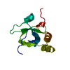
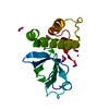
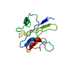
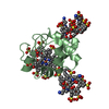
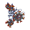
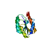
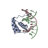
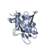
| Title | NMR structure of the N-terminal hath domain of human HDGF | ||||||
 Components Components | Hepatoma-derived growth factor | ||||||
 Keywords Keywords | HORMONE/GROWTH FACTOR / HDGF / hath domain / PWWP domain / heparin-binding / growth factor / HORMONE-GROWTH FACTOR COMPLEX | ||||||
| Function / homology |  Function and homology information Function and homology informationXBP1(S) activates chaperone genes / positive regulation of cell division / protein localization to nucleus / transcription repressor complex / tubulin binding / transcription corepressor binding / growth factor activity / DNA-binding transcription repressor activity, RNA polymerase II-specific / transcription corepressor activity / heparin binding ...XBP1(S) activates chaperone genes / positive regulation of cell division / protein localization to nucleus / transcription repressor complex / tubulin binding / transcription corepressor binding / growth factor activity / DNA-binding transcription repressor activity, RNA polymerase II-specific / transcription corepressor activity / heparin binding / actin binding / collagen-containing extracellular matrix / chromatin remodeling / RNA polymerase II cis-regulatory region sequence-specific DNA binding / nucleotide binding / negative regulation of transcription by RNA polymerase II / signal transduction / positive regulation of transcription by RNA polymerase II / extracellular space / RNA binding / extracellular region / nucleoplasm / nucleus / cytoplasmSimilarity search - Function | ||||||
| Biological species |  Homo sapiens (human) Homo sapiens (human) | ||||||
| Method | SOLUTION NMR / simulated annealing, molecular dynamics | ||||||
 Authors Authors | Sue, S.-C. / Chen, J.-Y. / Huang, T.-H. | ||||||
 Citation Citation | Journal: J.Mol.Biol. / Year: 2004 Title: Solution Structure and Heparin Interaction of Human Hepatoma-derived Growth Factor Authors: Sue, S.-C. / Chen, J.-Y. / Lee, S.-C. / Wu, W.-G. / Huang, T.-H. | ||||||
| History |
|
- Structure visualization
| Structure viewer | Molecule: MolmilJmol/JSmol |
|---|
- Downloads & links
-Download
| PDBx/mmCIF format | 1ri0.cif.gz | 620.7 KB | Display | PDBx/mmCIF format |
|---|---|---|---|---|
| PDB format | pdb1ri0.ent.gz | 537.4 KB | Display | PDB format |
| PDBx/mmJSON format | 1ri0.json.gz | Tree view | PDBx/mmJSON format | |
| Others |  Other downloads Other downloads |
-Validation report
| Arichive directory | https://data.pdbj.org/pub/pdb/validation_reports/ri/1ri0ftp://data.pdbj.org/pub/pdb/validation_reports/ri/1ri0 | HTTPS FTP |
|---|
-Related structure data
| Related structure data | |
|---|---|
| Similar structure data | |
| Other databases |
|


-Links
 PDBj
PDBj
- Assembly
| Deposited unit | 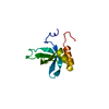
| |||||||||
|---|---|---|---|---|---|---|---|---|---|---|
| 1 |
| |||||||||
| NMR ensembles |
|
-Components
| #1: Protein | / The hath domain of hHDGF / HDGF / High-mobility group protein 1-like 2 / HMG-1L2 Mass: 12797.549 Da / Num. of mol.: 1 / Fragment: N-terminal 100 residues Source method: isolated from a genetically manipulated source Source: (gene. exp.) Homo sapiens (human) / Gene: HDGF / Plasmid: pET11d / Species (production host): Escherichia coli / Production host:  Escherichia coli BL21(DE3) (bacteria) / Strain (production host): BL21(DE3) / References: UniProt: P51858 Escherichia coli BL21(DE3) (bacteria) / Strain (production host): BL21(DE3) / References: UniProt: P51858 |
|---|
-Experimental details
-Experiment
| Experiment | Method: SOLUTION NMR | ||||||||||||||||
|---|---|---|---|---|---|---|---|---|---|---|---|---|---|---|---|---|---|
| NMR experiment |
| ||||||||||||||||
| NMR details | Text: The structure was determined by using standard triple-resonance NMR spectroscopy. |
- Sample preparation
| Details | Contents: 2 to 3mM 15N and 13C uniform labeled protein, 100mM phosphate buffer, 150mM NaCl, 1mM EDTA Solvent system: 95% H2O/5% D2O |
|---|---|
| Sample conditions | pH: 6.0 / Pressure: ambient / Temperature: 298 K |
-NMR measurement
| Radiation | Protocol: SINGLE WAVELENGTH / Monochromatic (M) / Laue (L): M | |||||||||||||||
|---|---|---|---|---|---|---|---|---|---|---|---|---|---|---|---|---|
| Radiation wavelength | Relative weight: 1 | |||||||||||||||
| NMR spectrometer |
|
- Processing
| NMR software |
| ||||||||||||||||||||
|---|---|---|---|---|---|---|---|---|---|---|---|---|---|---|---|---|---|---|---|---|---|
| Refinement | Method: simulated annealing, molecular dynamics / Software ordinal: 1 Details: The structures are based on a total of 1507 restraints, including 1245 NOE-derived distance constraints, 196 dihedral angle restraints, 66 distance restraints of hydrogen bonds. | ||||||||||||||||||||
| NMR representative | Selection criteria: fewest violations | ||||||||||||||||||||
| NMR ensemble | Conformer selection criteria: target function / Conformers calculated total number: 100 / Conformers submitted total number: 20 |
