 Movie
Movie Controller


+ Open data

- Basic information
| Entry | Database: PDB / ID: 1rhz | ||||||
|---|---|---|---|---|---|---|---|
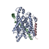
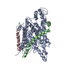
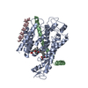
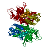
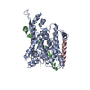
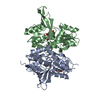
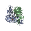
| Title | The structure of a protein conducting channel | ||||||
 Components Components |
| ||||||
 Keywords Keywords |  PROTEIN TRANSPORT / Protein translocation / SecY / Membrane Protein / Protein Channels PROTEIN TRANSPORT / Protein translocation / SecY / Membrane Protein / Protein Channels | ||||||
| Function / homology |  Function and homology informationintracellular protein transmembrane transport / SRP-dependent cotranslational protein targeting to membrane, translocation / signal sequence binding / protein transmembrane transporter activity / protein secretion / protein targeting / protein transport / plasma membrane Function and homology informationintracellular protein transmembrane transport / SRP-dependent cotranslational protein targeting to membrane, translocation / signal sequence binding / protein transmembrane transporter activity / protein secretion / protein targeting / protein transport / plasma membraneSimilarity search - Function | ||||||
| Biological species |   Methanocaldococcus jannaschii (archaea) Methanocaldococcus jannaschii (archaea) | ||||||
| Method | X-RAY DIFFRACTION / SYNCHROTRON / SAD / Resolution: 3.5 Å | ||||||
 Authors Authors | van den Berg, B. / Clemons Jr., W.M. / Collinson, I. / Modis, Y. / Hartmann, E. / Harrison, S.C. / Rapoport, T.A. | ||||||
 Citation Citation | Journal: Nature / Year: 2004 Title: X-ray structure of a protein-conducting channel. Authors: Van den Berg, B. / Clemons, W.M. / Collinson, I. / Modis, Y. / Hartmann, E. / Harrison, S.C. / Rapoport, T.A. | ||||||
| History |
| ||||||
| Remark 999 | sequence No suitable sequence database reference was available for chain C at the time of ...sequence No suitable sequence database reference was available for chain C at the time of processing this file. |
- Structure visualization
| Structure viewer | Molecule: MolmilJmol/JSmol |
|---|
- Downloads & links
-Download
| PDBx/mmCIF format | 1rhz.cif.gz | 104.8 KB | Display | PDBx/mmCIF format |
|---|---|---|---|---|
| PDB format | pdb1rhz.ent.gz | 81.6 KB | Display | PDB format |
| PDBx/mmJSON format | 1rhz.json.gz | Tree view | PDBx/mmJSON format | |
| Others |  Other downloads Other downloads |
-Validation report
| Arichive directory | https://data.pdbj.org/pub/pdb/validation_reports/rh/1rhzftp://data.pdbj.org/pub/pdb/validation_reports/rh/1rhz | HTTPS FTP |
|---|
-Related structure data


-Links
 PDBj
PDBj
- Assembly
| Deposited unit | 
| ||||||||
|---|---|---|---|---|---|---|---|---|---|
| 1 |
| ||||||||
| Unit cell |
|
-Components
| #1: Protein | Mass: 47482.891 Da / Num. of mol.: 1 Source method: isolated from a genetically manipulated source Source: (gene. exp.) Methanocaldococcus jannaschii (archaea)Gene: SECY, MJ0478 / Plasmid: pBAD22 / Production host:  Escherichia coli (E. coli) / Strain (production host): C43(DE3) / References: UniProt: Q60175 Escherichia coli (E. coli) / Strain (production host): C43(DE3) / References: UniProt: Q60175 |
|---|---|
| #2: Protein | Mass: 8451.144 Da / Num. of mol.: 1 Source method: isolated from a genetically manipulated source Source: (gene. exp.) Methanocaldococcus jannaschii (archaea)Gene: SECE, MJ0371 / Plasmid: pBAD22 / Production host: Escherichia coli (E. coli) / Strain (production host): C43(DE3) / References: UniProt: Q57817 |
| #3: Protein | Mass: 5967.010 Da / Num. of mol.: 1 Source method: isolated from a genetically manipulated source Source: (gene. exp.) Methanocaldococcus jannaschii (archaea)Plasmid: pBAD22 / Production host: Escherichia coli (E. coli) / Strain (production host): C43(DE3) / References: UniProt: P60460*PLUS |
-Experimental details
-Experiment
| Experiment | Method: X-RAY DIFFRACTION / Number of used crystals: 1 |
|---|
- Sample preparation
| Crystal | Density Matthews: 4.44 Å3/Da / Density % sol: 72.32 % | ||||||||||||||||||||||||
|---|---|---|---|---|---|---|---|---|---|---|---|---|---|---|---|---|---|---|---|---|---|---|---|---|---|
| Crystal grow | Temperature: 277 K / Method: vapor diffusion, hanging drop / pH: 9.5 Details: Peg 400, Glycine buffer, glycerol, sodium chloride, pH 9.5, VAPOR DIFFUSION, HANGING DROP, temperature 277K | ||||||||||||||||||||||||
| Crystal grow | *PLUS Temperature: 4 ℃ / Method: vapor diffusion, hanging drop / PH range low: 9.5 / PH range high: 9 | ||||||||||||||||||||||||
| Components of the solutions | *PLUS
|
-Data collection
| Diffraction | Mean temperature: 100 K |
|---|---|
| Diffraction source | Source: SYNCHROTRON / Site: NSLS  / Beamline: X25 / Wavelength: 0.9799 Å / Beamline: X25 / Wavelength: 0.9799 Å |
| Detector | Type: ADSC QUANTUM 4 / Detector: CCD / Date: Mar 1, 2002 |
| Radiation | Monochromator: Graphite / Protocol: SINGLE WAVELENGTH / Monochromatic (M) / Laue (L): M / Scattering type: x-ray |
| Radiation wavelength | Wavelength: 0.9799 Å / Relative weight: 1 |
| Reflection | Resolution: 3.5→10 Å / Num. all: 13799 / Num. obs: 13601 / % possible obs: 98.6 % / Observed criterion σ(F): 0 / Observed criterion σ(I): 0 / Rsym value: 0.08 / Net I/σ(I): 30.9 |
| Reflection shell | Resolution: 3.5→3.7 Å / Mean I/σ(I) obs: 2.74 / Num. unique all: 2010 / Rsym value: 0.67 / % possible all: 92.5 |
| Reflection | *PLUS Num. obs: 14439 / % possible obs: 98.4 % / Rmerge(I) obs: 0.08 |
| Reflection shell | *PLUS Highest resolution: 3.5 Å / Lowest resolution: 3.63 Å / % possible obs: 89.6 % / Num. unique obs: 1291 / Rmerge(I) obs: 0.67 |
- Processing
| Software |
| ||||||||||||||||||||||||||||||||||||||||||||||||||||||||||||
|---|---|---|---|---|---|---|---|---|---|---|---|---|---|---|---|---|---|---|---|---|---|---|---|---|---|---|---|---|---|---|---|---|---|---|---|---|---|---|---|---|---|---|---|---|---|---|---|---|---|---|---|---|---|---|---|---|---|---|---|---|---|
| Refinement | Method to determine structure: SAD / Resolution: 3.5→9.99 Å / Rfactor Rfree error: 0.013 / Data cutoff high absF: 845652.81 / Data cutoff high rms absF: 845652.81 / Data cutoff low absF: 0 / Isotropic thermal model: RESTRAINED / Cross valid method: THROUGHOUT / Stereochemistry target values: Engh & Huber
| ||||||||||||||||||||||||||||||||||||||||||||||||||||||||||||
| Solvent computation | Solvent model: FLAT MODEL / Bsol: 79.707 Å2 / ksol: 0.272543 e/Å3 | ||||||||||||||||||||||||||||||||||||||||||||||||||||||||||||
| Displacement parameters | Biso mean: 122.4 Å2
| ||||||||||||||||||||||||||||||||||||||||||||||||||||||||||||
| Refine analyze |
| ||||||||||||||||||||||||||||||||||||||||||||||||||||||||||||
| Refinement step | Cycle: LAST / Resolution: 3.5→9.99 Å
| ||||||||||||||||||||||||||||||||||||||||||||||||||||||||||||
| Refine LS restraints |
| ||||||||||||||||||||||||||||||||||||||||||||||||||||||||||||
| LS refinement shell | Resolution: 3.5→3.71 Å / Rfactor Rfree error: 0.047 / Total num. of bins used: 6
| ||||||||||||||||||||||||||||||||||||||||||||||||||||||||||||
| Xplor file | Serial no: 1 / Param file: PROTEIN_REP.PARAM / Topol file: PROTEIN.TOP | ||||||||||||||||||||||||||||||||||||||||||||||||||||||||||||
| Refinement | *PLUS Highest resolution: 3.5 Å / % reflection Rfree: 10 % / Rfactor Rfree: 0.334 | ||||||||||||||||||||||||||||||||||||||||||||||||||||||||||||
| Solvent computation | *PLUS | ||||||||||||||||||||||||||||||||||||||||||||||||||||||||||||
| Displacement parameters | *PLUS | ||||||||||||||||||||||||||||||||||||||||||||||||||||||||||||
| Refine LS restraints | *PLUS
| ||||||||||||||||||||||||||||||||||||||||||||||||||||||||||||
| LS refinement shell | *PLUS Lowest resolution: 3.63 Å |
