 Movie
Movie Controller


+ Open data

- Basic information
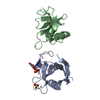
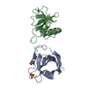
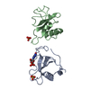
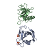
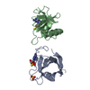
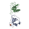
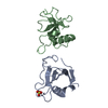
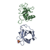
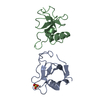
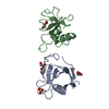
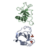
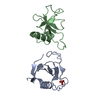
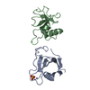
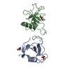
| Entry | Database: PDB / ID: 1rgg | ||||||
|---|---|---|---|---|---|---|---|
| Title | HYDROLASE, GUANYLORIBONUCLEASE | ||||||
 Components Components | RIBONUCLEASE | ||||||
 Keywords Keywords | HYDROLASE (GUANYLORIBONUCLEASE) | ||||||
| Function / homology |  Function and homology informationribonuclease T1 activity / ribonuclease T1 / RNA endonuclease activity / lyase activity / RNA binding / extracellular region Function and homology informationribonuclease T1 activity / ribonuclease T1 / RNA endonuclease activity / lyase activity / RNA binding / extracellular regionSimilarity search - Function | ||||||
| Biological species |  Streptomyces aureofaciens (bacteria) Streptomyces aureofaciens (bacteria) | ||||||
| Method | X-RAY DIFFRACTION / SYNCHROTRON / Resolution: 1.2 Å | ||||||
 Authors Authors | Sevcik, J. / Dauter, Z. / Lamzin, V.S. / Wilson, K.S. | ||||||
 Citation Citation | Journal: Acta Crystallogr.,Sect.D / Year: 1996 Title: Ribonuclease from Streptomyces aureofaciens at atomic resolution. Authors: Sevcik, J. / Dauter, Z. / Lamzin, V.S. / Wilson, K.S. #1: Journal: Acta Crystallogr.,Sect.D / Year: 1993Title: Complex of Ribonuclease from Streptomyces Aureofaciens with 2'-Gmp at 1.7A Resolution Authors: Sevcik, J. / Hill, C.P. / Dauter, Z. / Wilson, K.S. #2: Journal: Eur.J.Biochem. / Year: 1993Title: Complex of Ribonuclease Sa with a Cyclic Nucleotide and a Proposed Model for the Reaction Intermediate Authors: Sevcik, J. / Zegers, I. / Wyns, L. / Dauter, Z. / Wilson, K.S. #3: Journal: Acta Crystallogr.,Sect.B / Year: 1991Title: Determination and Restrained Least-Squares Refinement of the Structures of Ribonuclease Sa and its Complex with 3'-Guanylic Acid at 1.8 A Resolution Authors: Sevcik, J. / Dodson, E.J. / Dodson, G.G. #4: Journal: Trends Biochem.Sci. / Year: 1990Title: Comparison of Active Sites of Some Microbial Ribonucleases: Structural Basis for Guanylic Specificity Authors: Sevcik, J. / Sanishvili, R.G. / Pavlovsky, A.G. / Polyakov, K.M. #5: Journal: FEBS Lett. / Year: 1986Title: Amino Acid Sequence Determination of Guanyl-Specific Ribonuclease Sa from Streptomyces Aureofaciens Authors: Shlyapnikov, S.V. / Both, V. / Kulikov, V.A. / Dementiev, A.A. / Sevcik, J. / Zelinka, J. #6: Journal: Biochim.Biophys.Acta / Year: 1971Title: Exocellular Ribonuclease from Streptomyces Aureofaciens. I. Isolation and Purification Authors: Bacova, M. / Zelinkova, E. / Zelinka, J. #7: Journal: Biochim.Biophys.Acta / Year: 1971Title: Exocellular Ribonuclease from Streptomyces Aureofaciens. II. Properties and Specificity Authors: Zelinkova, E. / Bacova, M. / Zelinka, J. | ||||||
| History |
|
- Structure visualization
| Structure viewer | Molecule: MolmilJmol/JSmol |
|---|
- Downloads & links
-Download
| PDBx/mmCIF format | 1rgg.cif.gz | 144.5 KB | Display | PDBx/mmCIF format |
|---|---|---|---|---|
| PDB format | pdb1rgg.ent.gz | 119.2 KB | Display | PDB format |
| PDBx/mmJSON format | 1rgg.json.gz | Tree view | PDBx/mmJSON format | |
| Others |  Other downloads Other downloads |
-Validation report
| Arichive directory | https://data.pdbj.org/pub/pdb/validation_reports/rg/1rggftp://data.pdbj.org/pub/pdb/validation_reports/rg/1rgg | HTTPS FTP |
|---|
-Related structure data


-Links
 PDBj
PDBj- Assembly
| Deposited unit | 
| ||||||||
|---|---|---|---|---|---|---|---|---|---|
| 1 |
| ||||||||
| Unit cell |
| ||||||||
| Noncrystallographic symmetry (NCS) | NCS oper: (Code: given Matrix: (0.97091, 0.23576, 0.04183), Vector : |
-Components
| #1: Protein | Mass: 10582.492 Da / Num. of mol.: 2 / Source method: isolated from a natural source / Source: (natural) Streptomyces aureofaciens (bacteria) / References: UniProt: P05798, EC: 3.1.27.3#2: Chemical | Sulfate  Mass: 96.063 Da / Num. of mol.: 2 / Source method: obtained synthetically / Formula: SO4 Mass: 96.063 Da / Num. of mol.: 2 / Source method: obtained synthetically / Formula: SO4#3: Water | ChemComp-HOH / | Water Mass: 18.015 Da / Num. of mol.: 338 / Source method: isolated from a natural source / Formula: H2O Mass: 18.015 Da / Num. of mol.: 338 / Source method: isolated from a natural source / Formula: H2OCompound details | SECONDARY STRUCTURE BOUNDARIES HAVE BEEN DETERMINED USING SS PROGRAM (V.S.LAMZIN, EMBL HAMBURG) AS ...SECONDARY STRUCTURE BOUNDARIES | |
|---|
-Experimental details
-Experiment
| Experiment | Method: X-RAY DIFFRACTION |
|---|
- Sample preparation
| Crystal | Density Matthews: 2.34 Å3/Da / Density % sol: 47.45 % | ||||||||||||||||||||
|---|---|---|---|---|---|---|---|---|---|---|---|---|---|---|---|---|---|---|---|---|---|
| Crystal grow | *PLUS pH: 6.7 / Method: vapor diffusion | ||||||||||||||||||||
| Components of the solutions | *PLUS
|
-Data collection
| Diffraction source | Source: SYNCHROTRON / Site: EMBL/DESY, Hamburg  / Beamline: X11 / Wavelength: 0.92 / Beamline: X11 / Wavelength: 0.92 |
|---|---|
| Detector | Type: MARRESEARCH / Detector: IMAGE PLATE / Date: Nov 1, 1993 |
| Radiation | Monochromatic (M) / Laue (L): M / Scattering type: x-ray |
| Radiation wavelength | Wavelength: 0.92 Å / Relative weight: 1 |
| Reflection | Redundancy: 4.1 % / Rmerge(I) obs: 0.039 |
| Reflection | *PLUS Highest resolution: 1.2 Å / Num. obs: 60670 / % possible obs: 95.3 % / Num. measured all: 246637 / Biso Wilson estimate: 10.8 Å2 |
- Processing
| Software |
| |||||||||||||||||||||||||||||||||
|---|---|---|---|---|---|---|---|---|---|---|---|---|---|---|---|---|---|---|---|---|---|---|---|---|---|---|---|---|---|---|---|---|---|---|
| Refinement | Resolution: 1.2→10 Å / Num. parameters: 16942 / Num. restraintsaints: 20335 / σ(F): 0 / Stereochemistry target values: ENGH AND HUBER Details: ANISOU RECORDS CONTAIN ANISOTROPIC DISPLACEMENT PARAMETERS U11 U22 U33 U23 U13 U12 (ANGSTROMS**2) MULTIPLIED BY 10000. ISOTROPIC EQUIVALENTS OF ANISOTROPIC TEMPERATURE FACTORS ARE ALSO PRESENTED IN THIS ENTRY.
| |||||||||||||||||||||||||||||||||
| Solvent computation | Solvent model: BASED ON BABINET'S PRINCIPLE | |||||||||||||||||||||||||||||||||
| Refine analyze | Num. disordered residues: 11 / Occupancy sum non hydrogen: 1840 | |||||||||||||||||||||||||||||||||
| Refinement step | Cycle: LAST / Resolution: 1.2→10 Å
| |||||||||||||||||||||||||||||||||
| Refine LS restraints |
| |||||||||||||||||||||||||||||||||
| Software | *PLUS Name: SHELXL-96 / Classification: refinement | |||||||||||||||||||||||||||||||||
| Refine LS restraints | *PLUS
|
