 Movie
Movie Controller


+ Open data

- Basic information
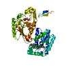
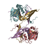
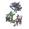
| Entry | Database: PDB / ID: 1r5o | ||||||
|---|---|---|---|---|---|---|---|
| Title | crystal structure analysis of sup35 complexed with GMPPNP | ||||||
 Components Components | Eukaryotic peptide chain release factor GTP-binding subunit | ||||||
 Keywords Keywords |  TRANSLATION / TRANSLATION TERMINATION / PEPTIDE RELEASE / GTPASE TRANSLATION / TRANSLATION TERMINATION / PEPTIDE RELEASE / GTPASE | ||||||
| Function / homology |  Function and homology information Function and homology informationEukaryotic Translation Termination / Nonsense Mediated Decay (NMD) independent of the Exon Junction Complex (EJC) / Nonsense Mediated Decay (NMD) enhanced by the Exon Junction Complex (EJC) / cytoplasmic translational termination / translation release factor complex / translation release factor activity / nuclear-transcribed mRNA catabolic process, deadenylation-dependent decay / Hydrolases; Acting on acid anhydrides; Acting on GTP to facilitate cellular and subcellular movement / translation / GTPase activity ...Eukaryotic Translation Termination / Nonsense Mediated Decay (NMD) independent of the Exon Junction Complex (EJC) / Nonsense Mediated Decay (NMD) enhanced by the Exon Junction Complex (EJC) / cytoplasmic translational termination / translation release factor complex / translation release factor activity / nuclear-transcribed mRNA catabolic process, deadenylation-dependent decay / Hydrolases; Acting on acid anhydrides; Acting on GTP to facilitate cellular and subcellular movement / translation / GTPase activity / GTP binding / cytosolSimilarity search - Function | ||||||
| Biological species |  Schizosaccharomyces pombe (fission yeast) Schizosaccharomyces pombe (fission yeast) | ||||||
| Method | X-RAY DIFFRACTION / SYNCHROTRON / MOLECULAR REPLACEMENT / Resolution: 3.2 Å | ||||||
 Authors Authors | Kong, C. / Song, H. | ||||||
 Citation Citation | Journal: Mol.Cell / Year: 2004 Title: Crystal structure and functional analysis of the eukaryotic class II release factor eRF3 from S. pombe Authors: Kong, C. / Ito, K. / Walsh, M.A. / Wada, M. / Liu, Y. / Kumar, S. / Barford, D. / Nakamura, Y. / Song, H. | ||||||
| History |
|
- Structure visualization
| Structure viewer | Molecule: MolmilJmol/JSmol |
|---|
- Downloads & links
-Download
| PDBx/mmCIF format | 1r5o.cif.gz | 98.2 KB | Display | PDBx/mmCIF format |
|---|---|---|---|---|
| PDB format | pdb1r5o.ent.gz | 73.3 KB | Display | PDB format |
| PDBx/mmJSON format | 1r5o.json.gz | Tree view | PDBx/mmJSON format | |
| Others |  Other downloads Other downloads |
-Validation report
| Arichive directory | https://data.pdbj.org/pub/pdb/validation_reports/r5/1r5oftp://data.pdbj.org/pub/pdb/validation_reports/r5/1r5o | HTTPS FTP |
|---|
-Related structure data
| Related structure data | 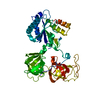 1r5bSC 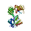 1r5nC S: Starting model for refinement C: citing same article ( |
|---|---|
| Similar structure data |


-Links
 PDBj
PDBj


- Assembly
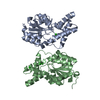
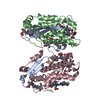
| Deposited unit | 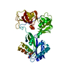
| ||||||||
|---|---|---|---|---|---|---|---|---|---|
| 1 |
| ||||||||
| Unit cell |
|
-Components
| #1: Protein | Mass: 52103.195 Da / Num. of mol.: 1 / Fragment: residues 196-662 Source method: isolated from a genetically manipulated source Source: (gene. exp.) Schizosaccharomyces pombe (fission yeast)Gene: sup35 / Plasmid: pGEX-6p-1 / Production host:  Escherichia coli (E. coli) / Strain (production host): Bl21-star / References: UniProt: O74718 Escherichia coli (E. coli) / Strain (production host): Bl21-star / References: UniProt: O74718 |
|---|---|
| #2: Chemical | ChemComp-GNP / 5'-Guanylyl imidodiphosphate  Mass: 522.196 Da / Num. of mol.: 1 / Source method: obtained synthetically / Formula: C10H17N6O13P3 Mass: 522.196 Da / Num. of mol.: 1 / Source method: obtained synthetically / Formula: C10H17N6O13P3Comment: GppNHp, GMPPNP, energy-carrying molecule analogue*YM |
| #3: Water | ChemComp-HOH / Water Mass: 18.015 Da / Num. of mol.: 56 / Source method: isolated from a natural source / Formula: H2O Mass: 18.015 Da / Num. of mol.: 56 / Source method: isolated from a natural source / Formula: H2O |
-Experimental details
-Experiment
| Experiment | Method: X-RAY DIFFRACTION / Number of used crystals: 1 |
|---|
- Sample preparation
| Crystal | Density Matthews: 3.09 Å3/Da / Density % sol: 59.85 % |
|---|---|
| Crystal grow | Temperature: 277 K / Method: vapor diffusion, hanging drop / pH: 7.5 Details: HEPES, Ethylene Glycol, PEG 8000, pH 7.5, VAPOR DIFFUSION, HANGING DROP, temperature 277K |
-Data collection
| Diffraction | Mean temperature: 100 K |
|---|---|
| Diffraction source | Source: SYNCHROTRON / Site: EMBL/DESY, HAMBURG  / Beamline: BW7A / Wavelength: 0.9724 Å / Beamline: BW7A / Wavelength: 0.9724 Å |
| Detector | Type: MARRESEARCH / Detector: CCD / Date: Jul 10, 2003 |
| Radiation | Monochromator: GRAPHITE / Protocol: SINGLE WAVELENGTH / Monochromatic (M) / Laue (L): M / Scattering type: x-ray |
| Radiation wavelength | Wavelength: 0.9724 Å / Relative weight: 1 |
| Reflection | Resolution: 3→20 Å / Num. all: 11985 / Num. obs: 11894 / % possible obs: 98.3 % / Observed criterion σ(I): 0 / Rmerge(I) obs: 0.14 / Rsym value: 0.14 / Net I/σ(I): 5.2 |
| Reflection shell | Resolution: 3→3.11 Å / Redundancy: 7.3 % / Rmerge(I) obs: 0.14 / Mean I/σ(I) obs: 0.7 / Num. unique all: 1134 / Rsym value: 0.14 / % possible all: 98.3 |
- Processing
| Software |
| ||||||||||||||||||||||||||||||||||||||||||||||||||||||||||||||||||||||||||||||||||||||||||||||||||||
|---|---|---|---|---|---|---|---|---|---|---|---|---|---|---|---|---|---|---|---|---|---|---|---|---|---|---|---|---|---|---|---|---|---|---|---|---|---|---|---|---|---|---|---|---|---|---|---|---|---|---|---|---|---|---|---|---|---|---|---|---|---|---|---|---|---|---|---|---|---|---|---|---|---|---|---|---|---|---|---|---|---|---|---|---|---|---|---|---|---|---|---|---|---|---|---|---|---|---|---|---|---|
| Refinement | Method to determine structure: MOLECULAR REPLACEMENT Starting model: 1R5B Resolution: 3.2→20 Å / Cor.coef. Fo:Fc: 0.877 / Cor.coef. Fo:Fc free: 0.813 / SU B: 23.886 / SU ML: 0.423 / Cross valid method: THROUGHOUT / ESU R Free: 0.621 / Stereochemistry target values: MAXIMUM LIKELIHOOD / Details: HYDROGENS HAVE BEEN ADDED IN THE RIDING POSITIONS
| ||||||||||||||||||||||||||||||||||||||||||||||||||||||||||||||||||||||||||||||||||||||||||||||||||||
| Solvent computation | Ion probe radii: 0.8 Å / Shrinkage radii: 0.8 Å / VDW probe radii: 1.4 Å / Solvent model: BABINET MODEL WITH MASK | ||||||||||||||||||||||||||||||||||||||||||||||||||||||||||||||||||||||||||||||||||||||||||||||||||||
| Displacement parameters | Biso mean: 48.353 Å2
| ||||||||||||||||||||||||||||||||||||||||||||||||||||||||||||||||||||||||||||||||||||||||||||||||||||
| Refinement step | Cycle: LAST / Resolution: 3.2→20 Å
| ||||||||||||||||||||||||||||||||||||||||||||||||||||||||||||||||||||||||||||||||||||||||||||||||||||
| Refine LS restraints |
| ||||||||||||||||||||||||||||||||||||||||||||||||||||||||||||||||||||||||||||||||||||||||||||||||||||
| LS refinement shell | Resolution: 3.2→3.281 Å / Total num. of bins used: 20 /
|
