 Movie
Movie Controller


+ Open data

- Basic information
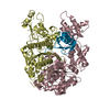
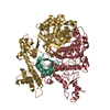
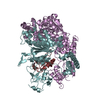
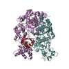
| Entry | Database: PDB / ID: 1r4n | ||||||
|---|---|---|---|---|---|---|---|
| Title | APPBP1-UBA3-NEDD8, an E1-ubiquitin-like protein complex with ATP | ||||||
 Components Components |
| ||||||
 Keywords Keywords |  CELL CYCLE CELL CYCLE | ||||||
| Function / homology |  Function and homology information Function and homology informationE1 NEDD8-activating enzyme / NEDD8 activating enzyme activity / endomitotic cell cycle / ubiquitin activating enzyme activity / NEDD8 transferase activity / regulation of proteolysis / mitotic DNA replication checkpoint signaling / protein neddylation / TGF-beta receptor signaling activates SMADs / anatomical structure morphogenesis ...E1 NEDD8-activating enzyme / NEDD8 activating enzyme activity / endomitotic cell cycle / ubiquitin activating enzyme activity / NEDD8 transferase activity / regulation of proteolysis / mitotic DNA replication checkpoint signaling / protein neddylation / TGF-beta receptor signaling activates SMADs / anatomical structure morphogenesis / regulation of neuron apoptotic process / post-translational protein modification / NIK-->noncanonical NF-kB signaling / Dectin-1 mediated noncanonical NF-kB signaling / Iron uptake and transport / protein modification process / modification-dependent protein catabolic process / protein localization / protein tag activity / UCH proteinases / Antigen processing: Ubiquitination & Proteasome degradation / Cargo recognition for clathrin-mediated endocytosis / Neddylation / ubiquitin-dependent protein catabolic process / neuron apoptotic process / regulation of apoptotic process / protein ubiquitination / regulation of cell cycle / protein heterodimerization activity / DNA damage response / ubiquitin protein ligase binding / regulation of transcription by RNA polymerase II / signal transduction / protein-containing complex / proteolysis / extracellular exosome / nucleoplasm / ATP binding / identical protein binding / nucleus / plasma membrane / cytosol / cytoplasmSimilarity search - Function | ||||||
| Biological species |  Homo sapiens (human) Homo sapiens (human) | ||||||
| Method | X-RAY DIFFRACTION / SYNCHROTRON / MOLECULAR REPLACEMENT / Resolution: 3.6 Å | ||||||
 Authors Authors | Walden, H. / Podgorski, M.S. / Holton, J.M. / Schulman, B.A. | ||||||
 Citation Citation | Journal: Mol.Cell / Year: 2003 Title: The structure of the APPBP1-UBA3-NEDD8-ATP complex reveals the basis for selective ubiquitin-like protein activation by an E1. Authors: Walden, H. / Podgorski, M.S. / Huang, D.T. / Miller, D.W. / Howard, R.J. / Minor, D.L. / Holton, J.M. / Schulman, B.A. | ||||||
| History |
| ||||||
| Remark 999 | SEQUENCE The residues 600-608, 700-706, 800-802 and 900-917 of the chains B,D,F,H were modelled ...SEQUENCE The residues 600-608, 700-706, 800-802 and 900-917 of the chains B,D,F,H were modelled originally as alanines due to poor electron density. The residue names were assigned arbitrarily and the connectivity is unclear. |
- Structure visualization
| Structure viewer | Molecule: MolmilJmol/JSmol |
|---|
- Downloads & links
-Download
| PDBx/mmCIF format | 1r4n.cif.gz | 726.4 KB | Display | PDBx/mmCIF format |
|---|---|---|---|---|
| PDB format | pdb1r4n.ent.gz | 594.5 KB | Display | PDB format |
| PDBx/mmJSON format | 1r4n.json.gz | Tree view | PDBx/mmJSON format | |
| Others |  Other downloads Other downloads |
-Validation report
| Arichive directory | https://data.pdbj.org/pub/pdb/validation_reports/r4/1r4nftp://data.pdbj.org/pub/pdb/validation_reports/r4/1r4n | HTTPS FTP |
|---|
-Related structure data
| Related structure data | 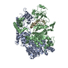 1r4mSC S: Starting model for refinement C: citing same article ( |
|---|---|
| Similar structure data |


-Links
 PDBj
PDBj


- Assembly
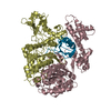
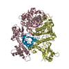
| Deposited unit | 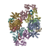
| ||||||||
|---|---|---|---|---|---|---|---|---|---|
| 1 | 
| ||||||||
| 2 | 
| ||||||||
| 3 | 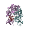
| ||||||||
| 4 | 
| ||||||||
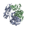
| Unit cell |
|
-Components
| #1: Protein | Mass: 59829.652 Da / Num. of mol.: 4 Source method: isolated from a genetically manipulated source Source: (gene. exp.) Homo sapiens (human) / Production host:  Escherichia coli (E. coli) / References: UniProt: Q13564 Escherichia coli (E. coli) / References: UniProt: Q13564#2: Protein | Mass: 48394.219 Da / Num. of mol.: 4 / Mutation: C216A Source method: isolated from a genetically manipulated source Source: (gene. exp.) Homo sapiens (human) / Production host: Escherichia coli (E. coli) / References: UniProt: Q8TBC4#3: Protein | Mass: 8573.978 Da / Num. of mol.: 4 Source method: isolated from a genetically manipulated source Source: (gene. exp.) Homo sapiens (human) / Gene: NEDD8 / Production host: Escherichia coli (E. coli) / References: UniProt: Q15843#4: Chemical | ChemComp-ATP / Adenosine triphosphate  Mass: 507.181 Da / Num. of mol.: 4 / Source method: obtained synthetically / Formula: C10H16N5O13P3 / Comment: ATP, energy-carrying molecule*YM Mass: 507.181 Da / Num. of mol.: 4 / Source method: obtained synthetically / Formula: C10H16N5O13P3 / Comment: ATP, energy-carrying molecule*YM#5: Chemical | ChemComp-ZN /   Mass: 65.409 Da / Num. of mol.: 4 / Source method: obtained synthetically / Formula: Zn Mass: 65.409 Da / Num. of mol.: 4 / Source method: obtained synthetically / Formula: Zn |
|---|
-Experimental details
-Experiment
| Experiment | Method: X-RAY DIFFRACTION / Number of used crystals: 1 |
|---|
- Sample preparation
| Crystal | Density Matthews: 3.02 Å3/Da / Density % sol: 59.21 % | ||||||||||||||||||||||||||||||||||||||||||||||||||||||||||||||||||||||||||||||||||||
|---|---|---|---|---|---|---|---|---|---|---|---|---|---|---|---|---|---|---|---|---|---|---|---|---|---|---|---|---|---|---|---|---|---|---|---|---|---|---|---|---|---|---|---|---|---|---|---|---|---|---|---|---|---|---|---|---|---|---|---|---|---|---|---|---|---|---|---|---|---|---|---|---|---|---|---|---|---|---|---|---|---|---|---|---|---|
| Crystal grow | Temperature: 291 K / Method: vapor diffusion, hanging drop / pH: 8 Details: peg10k, pH 8.0, VAPOR DIFFUSION, HANGING DROP, temperature 291K | ||||||||||||||||||||||||||||||||||||||||||||||||||||||||||||||||||||||||||||||||||||
| Crystal grow | *PLUS Temperature: 18 ℃ / pH: 7.6 / Method: vapor diffusion, hanging drop | ||||||||||||||||||||||||||||||||||||||||||||||||||||||||||||||||||||||||||||||||||||
| Components of the solutions | *PLUS
|
-Data collection
| Diffraction | Mean temperature: 200 K |
|---|---|
| Diffraction source | Source: SYNCHROTRON / Site: APS  / Beamline: 22-ID / Wavelength: 1 Å / Beamline: 22-ID / Wavelength: 1 Å |
| Detector | Type: MARRESEARCH / Detector: CCD / Date: Jul 7, 2003 |
| Radiation | Protocol: SINGLE WAVELENGTH / Monochromatic (M) / Laue (L): M / Scattering type: x-ray |
| Radiation wavelength | Wavelength: 1 Å / Relative weight: 1 |
| Reflection | Resolution: 3.6→50 Å / Num. obs: 64720 / % possible obs: 97.9 % / Observed criterion σ(F): 2 / Observed criterion σ(I): 1.9 / Redundancy: 41 % / Biso Wilson estimate: 69 Å2 / Rmerge(I) obs: 0.087 / Net I/σ(I): 12.6 |
| Reflection shell | Resolution: 3.6→3.7 Å / Rmerge(I) obs: 0.361 / Mean I/σ(I) obs: 1.9 / % possible all: 94.4 |
| Reflection | *PLUS Num. obs: 66111 / Num. measured all: 2710454 |
| Reflection shell | *PLUS % possible obs: 94.4 % |
- Processing
| Software |
| ||||||||||||||||||||||||||||||||||||||||||||||||||||||||||||
|---|---|---|---|---|---|---|---|---|---|---|---|---|---|---|---|---|---|---|---|---|---|---|---|---|---|---|---|---|---|---|---|---|---|---|---|---|---|---|---|---|---|---|---|---|---|---|---|---|---|---|---|---|---|---|---|---|---|---|---|---|---|
| Refinement | Method to determine structure: MOLECULAR REPLACEMENT Starting model: PDB entry 1R4M Resolution: 3.6→50 Å / σ(F): 0 / Stereochemistry target values: Engh & Huber
| ||||||||||||||||||||||||||||||||||||||||||||||||||||||||||||
| Displacement parameters | Biso mean: 92 Å2
| ||||||||||||||||||||||||||||||||||||||||||||||||||||||||||||
| Refinement step | Cycle: LAST / Resolution: 3.6→50 Å
| ||||||||||||||||||||||||||||||||||||||||||||||||||||||||||||
| Refine LS restraints |
| ||||||||||||||||||||||||||||||||||||||||||||||||||||||||||||
| Xplor file |
| ||||||||||||||||||||||||||||||||||||||||||||||||||||||||||||
| Refinement | *PLUS % reflection Rfree: 5 % / Rfactor Rfree: 0.29 | ||||||||||||||||||||||||||||||||||||||||||||||||||||||||||||
| Solvent computation | *PLUS | ||||||||||||||||||||||||||||||||||||||||||||||||||||||||||||
| Displacement parameters | *PLUS | ||||||||||||||||||||||||||||||||||||||||||||||||||||||||||||
| Refine LS restraints | *PLUS
|
