 Movie
Movie Controller


+ Open data

- Basic information
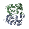
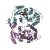
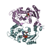
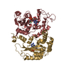
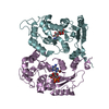
| Entry | Database: PDB / ID: 1r15 | ||||||
|---|---|---|---|---|---|---|---|
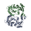
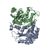
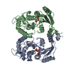
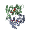
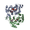
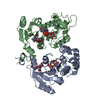
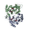
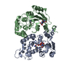
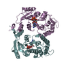
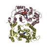
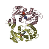
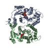
| Title | Aplysia ADP ribosyl cyclase with bound nicotinamide and R5P | ||||||
 Components Components | ADP-ribosyl cyclase | ||||||
 Keywords Keywords | HYDROLASE / ADP-ribosyl cyclase / cyclic ADP-ribose / NAADP / Ca2+ signalling | ||||||
| Function / homology |  Function and homology information Function and homology information2'-phospho-ADP-ribosyl cyclase/2'-phospho-cyclic-ADP-ribose transferase / NAD+ nucleotidase, cyclic ADP-ribose generating / NADP+ nucleosidase activity / Hydrolases; Glycosylases; Hydrolysing N-glycosyl compounds / single fertilization / transferase activity / cytoplasmic vesicle / membraneSimilarity search - Function | ||||||
| Biological species |  Aplysia californica (California sea hare) Aplysia californica (California sea hare) | ||||||
| Method | X-RAY DIFFRACTION / SYNCHROTRON / MOLECULAR REPLACEMENT / Resolution: 2.4 Å | ||||||
 Authors Authors | Love, M.L. / Szebenyi, D.M.E. / Kriksunov, I.A. / Thiel, D.J. / Munshi, C. / Graeff, R. / Lee, H.C. / Hao, Q. | ||||||
 Citation Citation | Journal: Structure / Year: 2004 Title: ADP-ribosyl cyclase; crystal structures reveal a covalent intermediate. Authors: Love, M.L. / Szebenyi, D.M. / Kriksunov, I.A. / Thiel, D.J. / Munshi, C. / Graeff, R. / Lee, H.C. / Hao, Q. | ||||||
| History |
| ||||||
| Remark 600 | HETEROGEN THE R5P UNDERGOES REACTION WITH THE PROTEIN TO FORM A 1-DEOXY INTERMEDIATE, LABELLED N, 1- ...HETEROGEN THE R5P UNDERGOES REACTION WITH THE PROTEIN TO FORM A 1-DEOXY INTERMEDIATE, LABELLED N, 1-DEOXY-RIBOFURANOSE-5'-PHOSPHATE. THE N IS COVALENTLY BOUND TO THE PROTEIN. |
- Structure visualization
| Structure viewer | Molecule: MolmilJmol/JSmol |
|---|
- Downloads & links
-Download
| PDBx/mmCIF format | 1r15.cif.gz | 394.4 KB | Display | PDBx/mmCIF format |
|---|---|---|---|---|
| PDB format | pdb1r15.ent.gz | 335.2 KB | Display | PDB format |
| PDBx/mmJSON format | 1r15.json.gz | Tree view | PDBx/mmJSON format | |
| Others |  Other downloads Other downloads |
-Validation report
| Arichive directory | https://data.pdbj.org/pub/pdb/validation_reports/r1/1r15ftp://data.pdbj.org/pub/pdb/validation_reports/r1/1r15 | HTTPS FTP |
|---|
-Related structure data


-Links
 PDBj
PDBj- Assembly
| Deposited unit | 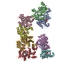
| ||||||||
|---|---|---|---|---|---|---|---|---|---|
| 1 | 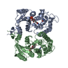
| ||||||||
| 2 | 
| ||||||||
| 3 | 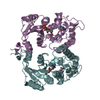
| ||||||||
| 4 | 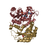
| ||||||||
| Unit cell |
|
-Components
| #1: Protein | / NAD+ / nucleosidase / NADASE / NAD glycohydrolase / ADRC Mass: 29579.945 Da / Num. of mol.: 8 Source method: isolated from a genetically manipulated source Source: (gene. exp.) Aplysia californica (California sea hare)Production host:  Escherichia coli (E. coli) / References: UniProt: P29241, NAD+ glycohydrolase Escherichia coli (E. coli) / References: UniProt: P29241, NAD+ glycohydrolase#2: Chemical | ChemComp-N /   Type: RNA linking / Mass: 214.110 Da / Num. of mol.: 8 / Source method: obtained synthetically / Formula: C5H11O7P Type: RNA linking / Mass: 214.110 Da / Num. of mol.: 8 / Source method: obtained synthetically / Formula: C5H11O7P#3: Chemical | ChemComp-NCA / Nicotinamide  Mass: 122.125 Da / Num. of mol.: 16 / Fragment: nicotinamide / Source method: obtained synthetically / Formula: C6H6N2O / Comment: medication*YM Mass: 122.125 Da / Num. of mol.: 16 / Fragment: nicotinamide / Source method: obtained synthetically / Formula: C6H6N2O / Comment: medication*YM |
|---|
-Experimental details
-Experiment
| Experiment | Method: X-RAY DIFFRACTION / Number of used crystals: 1 |
|---|
- Sample preparation
| Crystal | Density Matthews: 2.65 Å3/Da / Density % sol: 53.63 % | ||||||||||||||||||
|---|---|---|---|---|---|---|---|---|---|---|---|---|---|---|---|---|---|---|---|
| Crystal grow | Temperature: 316 K / Method: vapor diffusion, hanging drop / pH: 7.5 Details: 0.1 M Imidazole and 12-24 % PEG 4K, pH 7.5, VAPOR DIFFUSION, HANGING DROP, temperature 316K | ||||||||||||||||||
| Crystal grow | *PLUS Temperature: 18 ℃ / Method: vapor diffusion, hanging drop / Details: Munshi, C., (1999) J. Biol. Chem., 274, 30770. | ||||||||||||||||||
| Components of the solutions | *PLUS
|
-Data collection
| Diffraction | Mean temperature: 200 K |
|---|---|
| Diffraction source | Source: SYNCHROTRON / Site: CHESS  / Beamline: A1 / Wavelength: 0.919 Å / Beamline: A1 / Wavelength: 0.919 Å |
| Radiation | Protocol: SINGLE WAVELENGTH / Monochromatic (M) / Laue (L): M / Scattering type: x-ray |
| Radiation wavelength | Wavelength: 0.919 Å / Relative weight: 1 |
| Reflection | Resolution: 2.4→33 Å / Num. all: 64996 / Num. obs: 57197 / % possible obs: 88 % / Observed criterion σ(F): 0 / Observed criterion σ(I): 0 / Redundancy: 3.9 % / Rsym value: 0.108 |
| Reflection | *PLUS Rmerge(I) obs: 0.108 |
| Reflection shell | *PLUS Highest resolution: 2.4 Å / Lowest resolution: 2.6 Å / % possible obs: 44.7 % / Rmerge(I) obs: 0.31 |
- Processing
| Software |
| ||||||||||||||||||||
|---|---|---|---|---|---|---|---|---|---|---|---|---|---|---|---|---|---|---|---|---|---|
| Refinement | Method to determine structure: MOLECULAR REPLACEMENT / Resolution: 2.4→33 Å / σ(F): 0 / σ(I): 0 / Stereochemistry target values: Engh & Huber
| ||||||||||||||||||||
| Refinement step | Cycle: LAST / Resolution: 2.4→33 Å
| ||||||||||||||||||||
| Refine LS restraints |
| ||||||||||||||||||||
| Refinement | *PLUS Num. reflection obs: 57197 / Rfactor Rfree: 0.2694 / Rfactor Rwork: 0.2439 | ||||||||||||||||||||
| Solvent computation | *PLUS | ||||||||||||||||||||
| Displacement parameters | *PLUS |
