 Movie
Movie Controller

[English] 日本語
 Yorodumi
- PDB-1qy8: Crystal Structure of the N-domain of the ER Hsp90 chaperone GRP94... -
+ Open data

- Basic information
| Entry | Database: PDB / ID: 1qy8 | ||||||
|---|---|---|---|---|---|---|---|
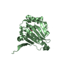
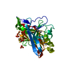
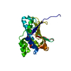
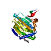
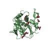
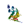
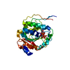
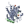
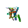
| Title | Crystal Structure of the N-domain of the ER Hsp90 chaperone GRP94 in complex with Radicicol | ||||||
 Components Components | Endoplasmin | ||||||
 Keywords Keywords |  CHAPERONE / GRP94 / gp96 / hsp90 / Radicicol CHAPERONE / GRP94 / gp96 / hsp90 / Radicicol | ||||||
| Function / homology |  Function and homology information Function and homology informationTrafficking and processing of endosomal TLR / Scavenging by Class A Receptors / Regulation of Insulin-like Growth Factor (IGF) transport and uptake by Insulin-like Growth Factor Binding Proteins (IGFBPs) / Interleukin-4 and Interleukin-13 signaling / Post-translational protein phosphorylation / sarcoplasmic reticulum lumen / : / ATP-dependent protein folding chaperone / melanosome / unfolded protein binding ...Trafficking and processing of endosomal TLR / Scavenging by Class A Receptors / Regulation of Insulin-like Growth Factor (IGF) transport and uptake by Insulin-like Growth Factor Binding Proteins (IGFBPs) / Interleukin-4 and Interleukin-13 signaling / Post-translational protein phosphorylation / sarcoplasmic reticulum lumen / : / ATP-dependent protein folding chaperone / melanosome / unfolded protein binding / protein folding / perinuclear region of cytoplasm / endoplasmic reticulum / ATP hydrolysis activity / ATP bindingSimilarity search - Function | ||||||
| Biological species |  Canis lupus familiaris (dog) Canis lupus familiaris (dog) | ||||||
| Method | X-RAY DIFFRACTION / SYNCHROTRON / MOLECULAR REPLACEMENT / Resolution: 1.85 Å | ||||||
 Authors Authors | Soldano, K.L. / Jivan, A. / Nicchitta, C.V. / Gewirth, D.T. | ||||||
 Citation Citation | Journal: J.Biol.Chem. / Year: 2003 Title: Structure of the N-terminal domain of GRP94. Basis for ligand specificity and regulation Authors: Soldano, K.L. / Jivan, A. / Nicchitta, C.V. / Gewirth, D.T. | ||||||
| History |
|
- Structure visualization
| Structure viewer | Molecule: MolmilJmol/JSmol |
|---|
- Downloads & links
-Download
| PDBx/mmCIF format | 1qy8.cif.gz | 63.6 KB | Display | PDBx/mmCIF format |
|---|---|---|---|---|
| PDB format | pdb1qy8.ent.gz | 45.1 KB | Display | PDB format |
| PDBx/mmJSON format | 1qy8.json.gz | Tree view | PDBx/mmJSON format | |
| Others |  Other downloads Other downloads |
-Validation report
| Arichive directory | https://data.pdbj.org/pub/pdb/validation_reports/qy/1qy8ftp://data.pdbj.org/pub/pdb/validation_reports/qy/1qy8 | HTTPS FTP |
|---|
-Related structure data
| Related structure data |  1qyeC  1u2oC  6d28C  1qy5 C: citing same article ( S: Starting model for refinement |
|---|---|
| Similar structure data |

-Links
 PDBj
PDBj


- Assembly
| Deposited unit | 
| ||||||||
|---|---|---|---|---|---|---|---|---|---|
| 1 |
| ||||||||
| Unit cell |
|
-Components
| #1: Protein | Mass: 30642.316 Da / Num. of mol.: 1 / Fragment: Residues 69-337 Source method: isolated from a genetically manipulated source Source: (gene. exp.) Canis lupus familiaris (dog) / Species: Canis lupus / Strain: familiaris / Gene: TRA1 / Plasmid: pGEXNB / Species (production host): Escherichia coli / Production host:  Escherichia coli BL21(DE3) (bacteria) / Strain (production host): BL21(DE3) / References: UniProt: P41148 Escherichia coli BL21(DE3) (bacteria) / Strain (production host): BL21(DE3) / References: UniProt: P41148 |
|---|---|
| #2: Chemical | ChemComp-RDI / Radicicol  Mass: 370.825 Da / Num. of mol.: 1 / Source method: obtained synthetically / Formula: C18H23ClO6 Mass: 370.825 Da / Num. of mol.: 1 / Source method: obtained synthetically / Formula: C18H23ClO6 |
| #3: Water | ChemComp-HOH / Water Mass: 18.015 Da / Num. of mol.: 169 / Source method: isolated from a natural source / Formula: H2O Mass: 18.015 Da / Num. of mol.: 169 / Source method: isolated from a natural source / Formula: H2O |
-Experimental details
-Experiment
| Experiment | Method: X-RAY DIFFRACTION / Number of used crystals: 1 |
|---|
- Sample preparation
| Crystal | Density Matthews: 2.23 Å3/Da / Density % sol: 44.73 % | ||||||||||||||||||||||||||||||||||||||||||
|---|---|---|---|---|---|---|---|---|---|---|---|---|---|---|---|---|---|---|---|---|---|---|---|---|---|---|---|---|---|---|---|---|---|---|---|---|---|---|---|---|---|---|---|
| Crystal grow | Temperature: 291 K / Method: vapor diffusion, hanging drop / pH: 7.6 Details: PEG 550 MME, Magnesium chloride, Tris, pH 7.6, VAPOR DIFFUSION, HANGING DROP, temperature 291K | ||||||||||||||||||||||||||||||||||||||||||
| Crystal grow | *PLUS Temperature: 18 ℃ / Method: vapor diffusion, hanging drop | ||||||||||||||||||||||||||||||||||||||||||
| Components of the solutions | *PLUS
|
-Data collection
| Diffraction | Mean temperature: 100 K |
|---|---|
| Diffraction source | Source: SYNCHROTRON / Site: APS  / Beamline: 19-BM / Wavelength: 1.0332 Å / Beamline: 19-BM / Wavelength: 1.0332 Å |
| Detector | Type: CUSTOM-MADE / Detector: CCD / Date: Sep 14, 2001 |
| Radiation | Monochromator: SAGITALLY FOCUSED Si(111) / Protocol: SINGLE WAVELENGTH / Monochromatic (M) / Laue (L): M / Scattering type: x-ray |
| Radiation wavelength | Wavelength: 1.0332 Å / Relative weight: 1 |
| Reflection | Resolution: 1.85→50 Å / Num. all: 23549 / Num. obs: 23549 / % possible obs: 99 % / Observed criterion σ(I): -3 / Redundancy: 7.3 % / Biso Wilson estimate: 25.8 Å2 / Rmerge(I) obs: 0.048 / Net I/σ(I): 36.3 |
| Reflection shell | Resolution: 1.85→1.92 Å / Rmerge(I) obs: 0.44 / Mean I/σ(I) obs: 1.89 / % possible all: 95.5 |
| Reflection | *PLUS Rmerge(I) obs: 0.055 |
| Reflection shell | *PLUS % possible obs: 95.5 % / Rmerge(I) obs: 0.44 |
- Processing
| Software |
| ||||||||||||||||||||||||||||||||||||
|---|---|---|---|---|---|---|---|---|---|---|---|---|---|---|---|---|---|---|---|---|---|---|---|---|---|---|---|---|---|---|---|---|---|---|---|---|---|
| Refinement | Method to determine structure: MOLECULAR REPLACEMENT Starting model: PDB entry 1QY5 1qy5 Resolution: 1.85→50 Å / Rfactor Rfree error: 0.006 / Data cutoff high absF: 694469.44 / Data cutoff high rms absF: 694469.44 / Data cutoff low absF: 0 / Isotropic thermal model: RESTRAINED / Cross valid method: THROUGHOUT / σ(F): 0 / Stereochemistry target values: Engh & Huber / Details: BULK SOLVENT MODEL USED
| ||||||||||||||||||||||||||||||||||||
| Solvent computation | Solvent model: FLAT MODEL / Bsol: 64.8282 Å2 / ksol: 0.372593 e/Å3 | ||||||||||||||||||||||||||||||||||||
| Displacement parameters | Biso mean: 43.6 Å2
| ||||||||||||||||||||||||||||||||||||
| Refine analyze |
| ||||||||||||||||||||||||||||||||||||
| Refinement step | Cycle: LAST / Resolution: 1.85→50 Å
| ||||||||||||||||||||||||||||||||||||
| Refine LS restraints |
| ||||||||||||||||||||||||||||||||||||
| LS refinement shell | Resolution: 1.85→1.92 Å / Rfactor Rfree error: 0.03 / Total num. of bins used: 10
| ||||||||||||||||||||||||||||||||||||
| Xplor file |
| ||||||||||||||||||||||||||||||||||||
| Refinement | *PLUS Lowest resolution: 6 Å / % reflection Rfree: 10 % / Rfactor Rfree: 0.277 | ||||||||||||||||||||||||||||||||||||
| Solvent computation | *PLUS | ||||||||||||||||||||||||||||||||||||
| Displacement parameters | *PLUS | ||||||||||||||||||||||||||||||||||||
| Refine LS restraints | *PLUS
| ||||||||||||||||||||||||||||||||||||
| LS refinement shell | *PLUS Rfactor Rfree: 0.269 / Rfactor Rwork: 0.214 |
