 Movie
Movie Controller

[English] 日本語
 Yorodumi
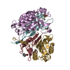
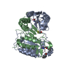
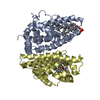
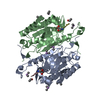
- PDB-1qtn: CRYSTAL STRUCTURE OF THE COMPLEX OF CASPASE-8 WITH THE TETRAPEPTI... -
+ Open data

- Basic information
| Entry | Database: PDB / ID: 1qtn | ||||||
|---|---|---|---|---|---|---|---|
| Title | CRYSTAL STRUCTURE OF THE COMPLEX OF CASPASE-8 WITH THE TETRAPEPTIDE INHIBITOR ACE-IETD-ALDEHYDE | ||||||
 Components Components |
| ||||||
 Keywords Keywords | HYDROLASE/HYDROLASE INHIBITOR /  APOPTOSIS / DITHIANE-DIOL / CASPASE / CYSTEINE-PROTEASE / HYDROLASE-HYDROLASE INHIBITOR COMPLEX APOPTOSIS / DITHIANE-DIOL / CASPASE / CYSTEINE-PROTEASE / HYDROLASE-HYDROLASE INHIBITOR COMPLEX | ||||||
| Function / homology |  Function and homology informationcaspase-8 / death effector domain binding / syncytiotrophoblast cell differentiation involved in labyrinthine layer development / FasL/ CD95L signaling / TRAIL signaling / CD95 death-inducing signaling complex / ripoptosome / Defective RIPK1-mediated regulated necrosis / Apoptotic execution phase / Activation, myristolyation of BID and translocation to mitochondria ...caspase-8 / death effector domain binding / syncytiotrophoblast cell differentiation involved in labyrinthine layer development / FasL/ CD95L signaling / TRAIL signaling / CD95 death-inducing signaling complex / ripoptosome / Defective RIPK1-mediated regulated necrosis / Apoptotic execution phase / Activation, myristolyation of BID and translocation to mitochondria / TRAIL-activated apoptotic signaling pathway / TRIF-mediated programmed cell death / TLR3-mediated TICAM1-dependent programmed cell death / Microbial modulation of RIPK1-mediated regulated necrosis / Regulation by c-FLIP / CASP8 activity is inhibited / Dimerization of procaspase-8 / Caspase activation via Death Receptors in the presence of ligand / positive regulation of macrophage differentiation / NF-kB activation through FADD/RIP-1 pathway mediated by caspase-8 and -10 / self proteolysis / response to cobalt ion / cysteine-type endopeptidase activity involved in apoptotic signaling pathway / death-inducing signaling complex / natural killer cell activation / negative regulation of necroptotic process / CLEC7A/inflammasome pathway / activation of cysteine-type endopeptidase activity / regulation of tumor necrosis factor-mediated signaling pathway / tumor necrosis factor receptor binding / death receptor binding / cysteine-type endopeptidase activity involved in apoptotic process / TNFR1-induced proapoptotic signaling / execution phase of apoptosis / regulation of innate immune response / RIPK1-mediated regulated necrosis / B cell activation / pyroptosis / Apoptotic cleavage of cellular proteins / positive regulation of proteolysis / macrophage differentiation / protein maturation / cellular response to organic cyclic compound / extrinsic apoptotic signaling pathway via death domain receptors / Caspase-mediated cleavage of cytoskeletal proteins / response to tumor necrosis factor / cysteine-type peptidase activity / negative regulation of canonical NF-kappaB signal transduction / extrinsic apoptotic signaling pathway / regulation of cytokine production / T cell activation / proteolysis involved in protein catabolic process / positive regulation of interleukin-1 beta production / Regulation of NF-kappa B signaling / apoptotic signaling pathway / Regulation of TNFR1 signaling / NOD1/2 Signaling Pathway / Regulation of necroptotic cell death / cellular response to mechanical stimulus / positive regulation of neuron apoptotic process / response to estradiol / lamellipodium / cell body / heart development / peptidase activity / scaffold protein binding / angiogenesis / positive regulation of canonical NF-kappaB signal transduction / response to ethanol / mitochondrial outer membrane / response to lipopolysaccharide / cytoskeleton / positive regulation of cell migration / positive regulation of apoptotic process / cysteine-type endopeptidase activity / apoptotic process / ubiquitin protein ligase binding / protein-containing complex binding / protein-containing complex / mitochondrion / proteolysis / nucleoplasm / identical protein binding / cytosol / cytoplasm Function and homology informationcaspase-8 / death effector domain binding / syncytiotrophoblast cell differentiation involved in labyrinthine layer development / FasL/ CD95L signaling / TRAIL signaling / CD95 death-inducing signaling complex / ripoptosome / Defective RIPK1-mediated regulated necrosis / Apoptotic execution phase / Activation, myristolyation of BID and translocation to mitochondria ...caspase-8 / death effector domain binding / syncytiotrophoblast cell differentiation involved in labyrinthine layer development / FasL/ CD95L signaling / TRAIL signaling / CD95 death-inducing signaling complex / ripoptosome / Defective RIPK1-mediated regulated necrosis / Apoptotic execution phase / Activation, myristolyation of BID and translocation to mitochondria / TRAIL-activated apoptotic signaling pathway / TRIF-mediated programmed cell death / TLR3-mediated TICAM1-dependent programmed cell death / Microbial modulation of RIPK1-mediated regulated necrosis / Regulation by c-FLIP / CASP8 activity is inhibited / Dimerization of procaspase-8 / Caspase activation via Death Receptors in the presence of ligand / positive regulation of macrophage differentiation / NF-kB activation through FADD/RIP-1 pathway mediated by caspase-8 and -10 / self proteolysis / response to cobalt ion / cysteine-type endopeptidase activity involved in apoptotic signaling pathway / death-inducing signaling complex / natural killer cell activation / negative regulation of necroptotic process / CLEC7A/inflammasome pathway / activation of cysteine-type endopeptidase activity / regulation of tumor necrosis factor-mediated signaling pathway / tumor necrosis factor receptor binding / death receptor binding / cysteine-type endopeptidase activity involved in apoptotic process / TNFR1-induced proapoptotic signaling / execution phase of apoptosis / regulation of innate immune response / RIPK1-mediated regulated necrosis / B cell activation / pyroptosis / Apoptotic cleavage of cellular proteins / positive regulation of proteolysis / macrophage differentiation / protein maturation / cellular response to organic cyclic compound / extrinsic apoptotic signaling pathway via death domain receptors / Caspase-mediated cleavage of cytoskeletal proteins / response to tumor necrosis factor / cysteine-type peptidase activity / negative regulation of canonical NF-kappaB signal transduction / extrinsic apoptotic signaling pathway / regulation of cytokine production / T cell activation / proteolysis involved in protein catabolic process / positive regulation of interleukin-1 beta production / Regulation of NF-kappa B signaling / apoptotic signaling pathway / Regulation of TNFR1 signaling / NOD1/2 Signaling Pathway / Regulation of necroptotic cell death / cellular response to mechanical stimulus / positive regulation of neuron apoptotic process / response to estradiol / lamellipodium / cell body / heart development / peptidase activity / scaffold protein binding / angiogenesis / positive regulation of canonical NF-kappaB signal transduction / response to ethanol / mitochondrial outer membrane / response to lipopolysaccharide / cytoskeleton / positive regulation of cell migration / positive regulation of apoptotic process / cysteine-type endopeptidase activity / apoptotic process / ubiquitin protein ligase binding / protein-containing complex binding / protein-containing complex / mitochondrion / proteolysis / nucleoplasm / identical protein binding / cytosol / cytoplasmSimilarity search - Function | ||||||
| Biological species |  Homo sapiens (human) Homo sapiens (human) | ||||||
| Method | X-RAY DIFFRACTION / SYNCHROTRON / molecular replacement / Resolution: 1.2 Å | ||||||
 Authors Authors | Watt, W. / Watenpaugh, K.D. | ||||||
 Citation Citation | Journal: Structure Fold.Des. / Year: 1999 Title: The atomic-resolution structure of human caspase-8, a key activator of apoptosis. Authors: Watt, W. / Koeplinger, K.A. / Mildner, A.M. / Heinrikson, R.L. / Tomasselli, A.G. / Watenpaugh, K.D. | ||||||
| History |
|
- Structure visualization
| Structure viewer | Molecule: MolmilJmol/JSmol |
|---|
- Downloads & links
-Download
| PDBx/mmCIF format | 1qtn.cif.gz | 73.4 KB | Display | PDBx/mmCIF format |
|---|---|---|---|---|
| PDB format | pdb1qtn.ent.gz | 52 KB | Display | PDB format |
| PDBx/mmJSON format | 1qtn.json.gz | Tree view | PDBx/mmJSON format | |
| Others |  Other downloads Other downloads |
-Validation report
| Arichive directory | https://data.pdbj.org/pub/pdb/validation_reports/qt/1qtnftp://data.pdbj.org/pub/pdb/validation_reports/qt/1qtn | HTTPS FTP |
|---|
-Related structure data
| Related structure data |  1cp3S S: Starting model for refinement |
|---|---|
| Similar structure data |


-Links
 PDBj
PDBj


- Assembly
| Deposited unit | 
| ||||||||
|---|---|---|---|---|---|---|---|---|---|
| 1 | 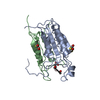
| ||||||||
| Unit cell |
| ||||||||
| Components on special symmetry positions |
|
-Components
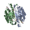
| #1: Protein | Caspase 8 Mass: 18640.119 Da / Num. of mol.: 1 / Fragment: P18 FRAGMENT Source method: isolated from a genetically manipulated source Source: (gene. exp.) Homo sapiens (human) / Production host:  Escherichia coli (E. coli) Escherichia coli (E. coli)References: UniProt: Q14790, Hydrolases; Acting on peptide bonds (peptidases); Cysteine endopeptidases | ||||
|---|---|---|---|---|---|
| #2: Protein | Caspase 8 Mass: 10901.364 Da / Num. of mol.: 1 / Fragment: P11 FRAGMENT Source method: isolated from a genetically manipulated source Source: (gene. exp.) Homo sapiens (human) / Production host: Escherichia coli (E. coli)References: UniProt: Q14790, Hydrolases; Acting on peptide bonds (peptidases); Cysteine endopeptidases | ||||
| #3: Protein/peptide | 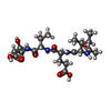  Type: Peptide-like / Class: Inhibitor / Mass: 488.532 Da / Num. of mol.: 1 / Source method: obtained synthetically / Details: CHEMICALLY SYNTHESIZED Type: Peptide-like / Class: Inhibitor / Mass: 488.532 Da / Num. of mol.: 1 / Source method: obtained synthetically / Details: CHEMICALLY SYNTHESIZEDReferences: N-(2-oxoethyl)-L-isoleucyl-L-alpha-glutamyl-N-[(1R)-2-carboxy-1-formylethyl]-L-threoninamide | ||||
| #4: Chemical | 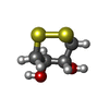  Mass: 152.235 Da / Num. of mol.: 2 / Source method: obtained synthetically / Formula: C4H8O2S2 Mass: 152.235 Da / Num. of mol.: 2 / Source method: obtained synthetically / Formula: C4H8O2S2#5: Water | ChemComp-HOH / | Water Mass: 18.015 Da / Num. of mol.: 332 / Source method: isolated from a natural source / Formula: H2O Mass: 18.015 Da / Num. of mol.: 332 / Source method: isolated from a natural source / Formula: H2OCompound details | THERE IS A COVALENT BOND BETWEEN ATOM SG OF CYS360A AND ATOM C OF ASJ504C, FORMING A THIOHEMIAC | |
-Experimental details
-Experiment
| Experiment | Method: X-RAY DIFFRACTION / Number of used crystals: 1 |
|---|
- Sample preparation
| Crystal | Density Matthews: 2.42 Å3/Da / Density % sol: 49.13 % | ||||||||||||||||||||||||||||||
|---|---|---|---|---|---|---|---|---|---|---|---|---|---|---|---|---|---|---|---|---|---|---|---|---|---|---|---|---|---|---|---|
| Crystal grow | Temperature: 282 K / Method: vapor diffusion, sitting drop / pH: 8 Details: 3 MICROLITERS PROTEIN (8.4 MG/ ML IN 20 MM TRISHCL, 100MM DTT AT PH 8.0) MIXED WITH 3 MICROLITERS WELL BUFFER (1.4 SODIUM CITRATE, 0.1M HEPES, AT PH 8.0) AT 4 DEG. C, VAPOR DIFFUSION, ...Details: 3 MICROLITERS PROTEIN (8.4 MG/ ML IN 20 MM TRISHCL, 100MM DTT AT PH 8.0) MIXED WITH 3 MICROLITERS WELL BUFFER (1.4 SODIUM CITRATE, 0.1M HEPES, AT PH 8.0) AT 4 DEG. C, VAPOR DIFFUSION, SITTING DROP, temperature 282K | ||||||||||||||||||||||||||||||
| Crystal grow | *PLUS Temperature: 4 ℃ | ||||||||||||||||||||||||||||||
| Components of the solutions | *PLUS
|
-Data collection
| Diffraction | Mean temperature: 105 K |
|---|---|
| Diffraction source | Source: SYNCHROTRON / Site: APS  / Beamline: 17-ID / Wavelength: 1.03 / Beamline: 17-ID / Wavelength: 1.03 |
| Detector | Type: BRUKER / Detector: CCD / Date: Sep 9, 1998 / Details: NON-FOCUSING |
| Radiation | Monochromator: SILICON / Protocol: SINGLE WAVELENGTH / Monochromatic (M) / Laue (L): M / Scattering type: x-ray |
| Radiation wavelength | Wavelength: 1.03 Å / Relative weight: 1 |
| Reflection | Resolution: 1.2→20 Å / Num. obs: 79890 / % possible obs: 87 % / Observed criterion σ(I): 0 / Redundancy: 3.73 % / Rmerge(I) obs: 0.068 |
| Reflection shell | Resolution: 1.2→1.24 Å / Rmerge(I) obs: 0.26 / Mean I/σ(I) obs: 2.35 / % possible all: 82 |
| Reflection | *PLUS Num. measured all: 298890 |
- Processing
| Software |
| |||||||||||||||||||||||||||||||||
|---|---|---|---|---|---|---|---|---|---|---|---|---|---|---|---|---|---|---|---|---|---|---|---|---|---|---|---|---|---|---|---|---|---|---|
| Refinement | Method to determine structure: molecular replacement Starting model: 1CP3 Resolution: 1.2→8 Å / σ(F): 4 / Stereochemistry target values: ENGH & HUBER
| |||||||||||||||||||||||||||||||||
| Solvent computation | Solvent model: SWAT | |||||||||||||||||||||||||||||||||
| Refinement step | Cycle: LAST / Resolution: 1.2→8 Å
| |||||||||||||||||||||||||||||||||
| Refine LS restraints |
| |||||||||||||||||||||||||||||||||
| Software | *PLUS Name: SHELXL-97 / Classification: refinement | |||||||||||||||||||||||||||||||||
| Refinement | *PLUS σ(F): 4 / % reflection Rfree: 5.3 % / Rfactor obs: 0.143 | |||||||||||||||||||||||||||||||||
| Solvent computation | *PLUS | |||||||||||||||||||||||||||||||||
| Displacement parameters | *PLUS | |||||||||||||||||||||||||||||||||
| Refine LS restraints | *PLUS
|
